
Presentation
Chronic headaches, no focal neurological symptoms. A routine check-up unexpectedly detected abnormalities
Patient Data


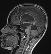
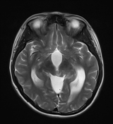
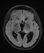
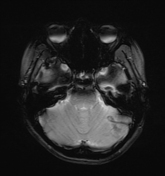
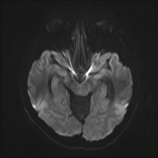

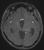
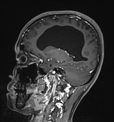
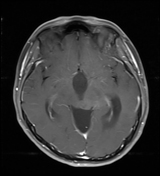
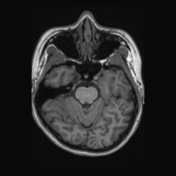
On the MRI of the brain with contrast enhancement, the left side of the tectal plate is noted to be enlarged by a nodular mass measuring approximately 16 x 11 x 13 mm. It shows increased signal on T2W and FLAIR, low signal on T1, and does not demonstrate contrast enhancement or restricted diffusion. The lesion extends into the left thalamic region, where a small nodule shows intrinsic high signal on T1W, MRA, as well as T1FS +C, no hypointense on GRE, no restricted diffusion.
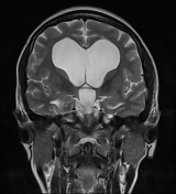
The aqueduct is compressed with prominent obstructive hydrocephalus of the third ventricle and lateral ventricle, with an Evan's index of approximately 0.53.
There is no transependymal edema, and the corpus callosum is markedly thinned.
The remainder of the brain shows no significant findings, with no abnormalities noted in or outside the axis.
Case Discussion
The imaging findings suggest that this could be a tectal glioma complicated by obstructive hydrocephalus due to compression of the aqueduct.
However, the lesion extends predominantly into the thalamic region, which necessitates a differential diagnosis with a thalamic glioma (usually high grade), whereas tectal gliomas are usually indolent.
A hyperintense nodule on T1, MRA, as well as T1FS +C, could represent lesions that shorten T1 relaxation time. Lesions such as small foci of melanin, fat, or hemorrhage are unlikely. It is more likely a form of mineralization, not necessarily coarse calcification. Not all mineralizations appear dark on SWI, and certainly not on GRE images with such low sensitivity. Although the presence of this nodule may not be highly significant, it slightly weakens the certainty of a diagnosis of low-grade indolent tectal glioma.
The patient did not undergo any further interventions and was discharged. The patient returned for follow-up at our hospital twice and underwent MRI, which showed that the lesions remained almost unchanged compared to previous examinations


 Unable to process the form. Check for errors and try again.