
Presentation
Pregnant lady with acute right lower abdominal pain.
Patient Data


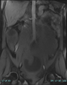
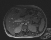
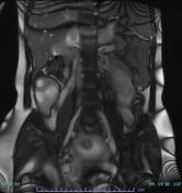
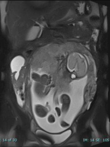
The right ovary is enlarged, measuring about (9 x 5 x 10 cm) with evidence of multiple cystic structures and surrounding free fluid.
The ovarian parenchyma appears heterogeneous with mild increase in signal intensity on T2WI without hyperintense foci on T1WI.
The right ovarian vein appears engorged.
The left ovary appears normal in size.
The gravid uterus shows a single fetus.
Case Discussion
This patient presented with acute right lower abdominal pain at 32 weeks of gestation. The ultrasound examination was inconclusive.
On unenhanced pelvic MRI, the right ovary appears high in position (displaced by the gravid uterus), enlarged and oedematous, associated with cystic changes, engorged ovarian vein and surrounding free fluid. In the sitting of the clinical presentation, findings suggests the diagnosis of ovarian torsion.
In surgery, the right ovary and fallopian tube were dusky and were resected.
Pathology report reveled oedematous haemorrhagic changes of the ovarian stroma, with cystic spaces and microcalcifications, consistent with torsion.
No evidence of tumour.


 Unable to process the form. Check for errors and try again.