
Presentation
Headache.
Patient Data
Age: 40 years
From the case:
Thrombosed berry aneurysm

Download
Info

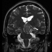
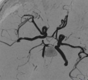
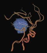
Partially thrombosed left MCA berry aneurysm is seen as a heterogeneous rounded structure with blood products resulting in a signal drop. The MRA shows the non-thrombosed part of the aneurysm located in the communicant posterior cerebral artery causing mass effect on the midbrain.
Download
Info
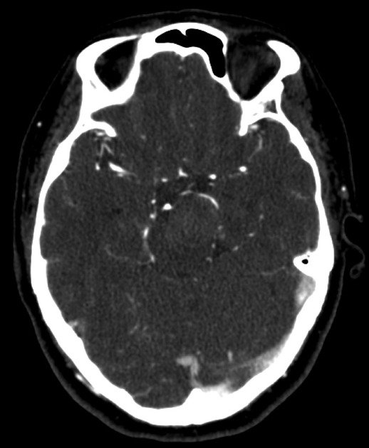
A CT performed 4 months after, shows a decrease in size.


 Unable to process the form. Check for errors and try again.