
Presentation
This middle-aged female patient presented with hip pain.
Patient Data
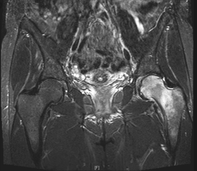
Coronal short-tau inversion recovery (STIR) MR image shows increased signal (bone oedema) within the left femoral head, neck and proximal metaphysis. There is also a small hip joint effusion.
Case Discussion
A follow-up study at 3 months shows resolution of most of the bone oedema. Transient osteoporosis is a self-limiting disease of unknown aetiology. It occurs in middle-aged males and females in late pregnancy typically. The hip is the most common joint affected. There is rapid-onset hip pain associated with demineralisation, usually unilateral. MRI findings are of diffuse marrow oedema and a small joint effusion. Bone scans show increased uptake. Spontaneous recovery occurs within 2-6 months. The differential diagnosis includes avascular necrosis, metastases, and reflex sympathetic dystrophy. Evidence of subchondral bone collapse, serpiginous subchondral interface, or cold spots on bone scan should prompt a diagnosis of AVN rather than transient osteoporosis.


 Unable to process the form. Check for errors and try again.