
Patient Data

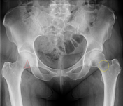
Unremarkable pelvic appearances.
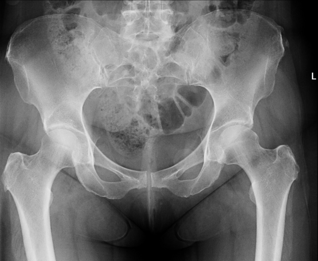
Red triangle on the right shows Babcock's triangle.
Yellow circle on the left shows Ward's triangle.
Case Discussion
This pelvic radiograph nicely shows two characteristic anatomical features of the proximal femur, each formed by the surrounding structural bony trabeculae. The lucent areas are important to recognise as "normal" features of the bony anatomy in this region to avoid the overcalling of false positive pathology.
Babcock's triangle is a fairly lucent focus found in the subcapital femoral neck which may be seen on anteroposterior radiographs of the hip. It is pertinent to be aware that historically this was often the region in which tuberculosis of the hip joint started.
Ward's triangle is a radiolucent area between different groups of trabeculae in the neck of femur (specifically the principal compressive, secondary compressive and primary tensile trabeculae). Its importance to radiologists lies in its use as the site to measure bone mineral density when performing DEXA assessment. It is usually the femoral neck region with the lowest bone mineral density 1.


 Unable to process the form. Check for errors and try again.