
Presentation
Headache.
Patient Data
Age: 50 years
Gender: Female
From the case:
Trichilemmal cyst


Download
Info
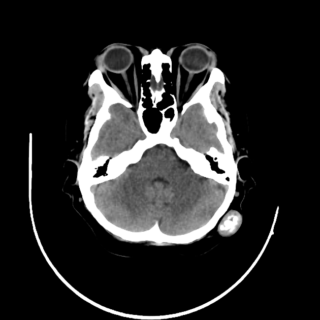
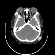
Non-contrast CT scan of the brain showed no intracranial pathology.
Incidentally noted left occipital scalp subcutaneous lesion showing coarse calcification. Intact underlying calvarium. A diagnosis of trichilemmal cyst was made.
Case Discussion
Trichilemmal cysts (TC) are keratin-filled cysts with a wall resembling the external root sheath of a hair follicle. They are the most common subcutaneous cysts of the scalp.
On imaging, these lesions can be either a cystic or solid mass which may show internal foci of calcifications.


 Unable to process the form. Check for errors and try again.