
Presentation
Primary infertility.
Patient Data
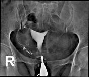
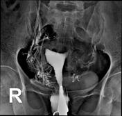
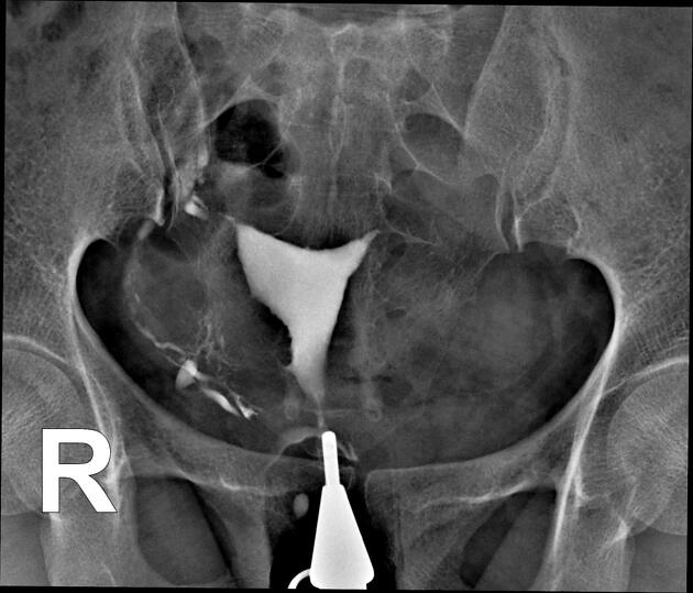
After the injection of contrast through the cervical canal, the uterine cavity becomes filled.
The body of the uterus is located in the midline of the pelvic cavity and displays a normal shape and contour.
Both fallopian tubes were not opacified from the primary segment of the isthmus region due to tubal occlusion.
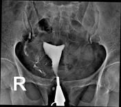
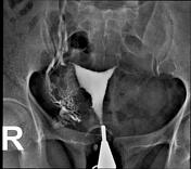
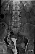
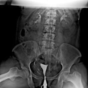
Following the administration of more contrast, there was an intravasation of the contrast media into the myometrial and right gonadal veins, which subsequently entered the renal veins.
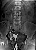
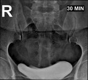
After three minutes of starting the procedure, the contrast material entered the kidneys, filling the pelvicalyceal system, and then flowed into the ureters and urinary bladder, creating an appearance similar to an intravenous pyelogram (IVP).
Case Discussion
IVP-like appearance during the HSG procedure indicates a possible tubal occlusion due to resistance to the flow of contrast material into the fallopian tubes and subsequently into the gonadal veins.


 Unable to process the form. Check for errors and try again.