
Presentation
Primary infertility.
Patient Data
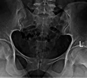
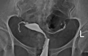

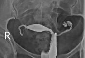

After contrast injection through the cervical canal, the uterine cavity and both fallopian tubes are filled.
The body of the uterus is located in the right paramidline of the pelvic cavity and has a normal shape and contour.
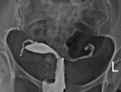
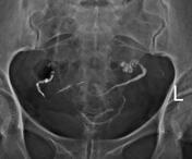
Both fallopian tubes are filled, demonstrating normal length, but they have abnormal peristalsis with retained contrast material without spillage, which suggests tubal occlusion.
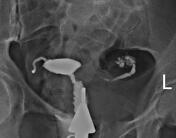
Small diverticular-like outpouchings in the ampullary region of the left fallopian tube, giving a rosette-like appearance.
Case Discussion
Hysterosalpingogram (HSG) findings indicated that bilateral tubal occlusion with a rosette-like appearance on the left side. The features of HSG, particularly the rosette-like appearance, strongly suggest a sequel to TB salpingitis1.


 Unable to process the form. Check for errors and try again.