
Patient Data
Age: Adult
From the case:
Tuberculous abscess
Download
Info
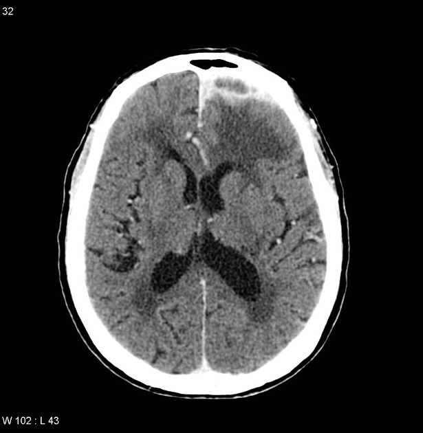
Peripherally enhancing extra-axial left frontal lesion. Significant adjacent vasogenic edema in the left cerebral hemisphere with partial effacement of the lateral ventricle. More superiorly in the frontal lobe is an enhancing nodule.
Case Discussion
The patient went on to have an evacuation of the collection.
Histology
Sections show a large area of caseating granulomatous inflammation associated with a fibrous type capsule, located superficially within the section of cerebral cortex. The ZN-stained sections confirm the presence of some scattered acid-fast bacilli.
Identification of mycobacteria on microscopy is usually seen to indicate a tuberculous abscess rather than tuberculoma.


 Unable to process the form. Check for errors and try again.