
Presentation
Admitted to the pulmonology department for pulmonary tuberculosis. Complaints of a headache.
Patient Data


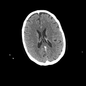
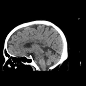
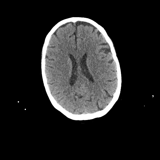
enlargement of the temporal horn of the right lateral ventricle
hypodensity around the trigone of the lateral ventricle representing vasogenic oedema
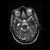
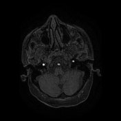
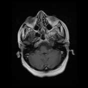
mild enhancement of the choroid plexus on enhanced CT
the left choroid plexus is unremarkable

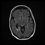
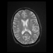
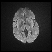
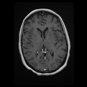
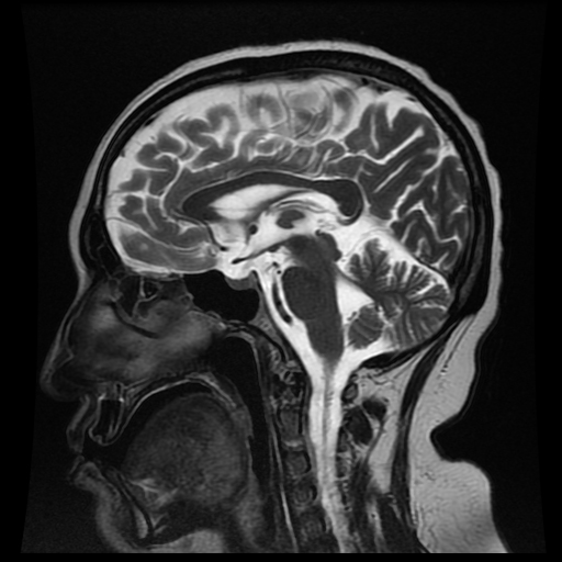
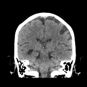
enlargement of the temporal horn of the right lateral ventricle with associated vasogenic oedema around the trigone
mild enhancement of the right choroid plexus of the lateral ventricle
no obvious leptomeningeal enhancement

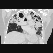
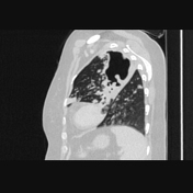
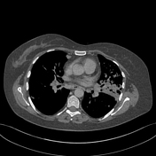
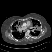
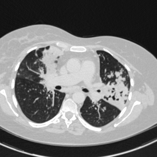
There are multiple alveolar consolidations, cavitations, tree-in-bud patterns, and centrilobular nodules at the upper lobes and left lower lobe.
Case Discussion
CT and brain MRI features of choroid plexitis with associated ventriculitis. This patient's history and chest CT are highly suggestive of the tuberculous origin of this choroid plexitis.


 Unable to process the form. Check for errors and try again.