
Presentation
Left knee - non-traumatic pain, swelling, restricted movements for about 5 months. Difficulty in walking - 2 weeks.
Patient Data

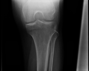
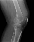
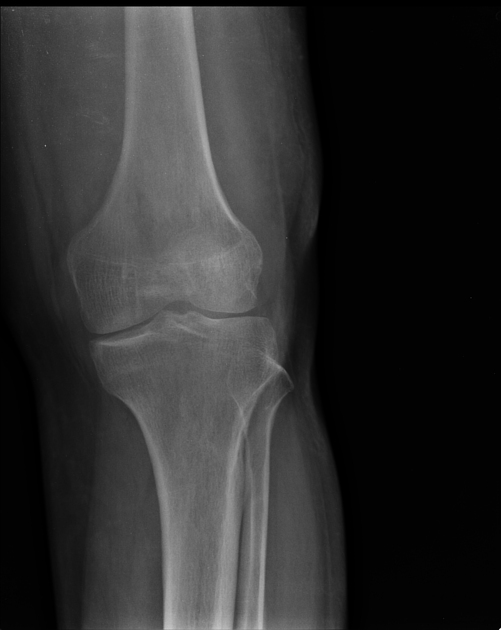
Periarticular osteopenia. Supra and infrapatellar soft tissue density. Widened intercondylar notch.
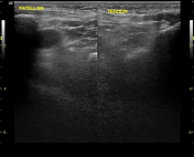
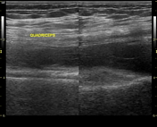


Diffuse synovial thickening with hypervascularity was seen involving suprapatellar, infrapatellar, medial, lateral joint recesses, and also in posterior joint space.
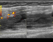
Collection / granulation was noted extending cranially to a suprapatellar pouch, into muscle.
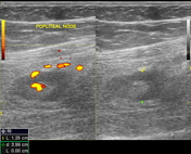
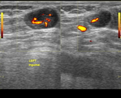
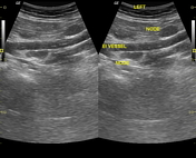
Lymph nodes are noted in the left iliac - inguinal - popliteal regions.
Case Discussion
Knee arthroscopy was done. Defect was seen in the suprapatellar capsule which explains collection outside joint capsular in the distal thigh.
Synovitis is a non-specific finding. However, extra-articular collection and lymphadenopathy favor infective etiology rather than inflammatory like rheumatoid.
Histopathology - Tuberculosis.


 Unable to process the form. Check for errors and try again.