
Presentation
Pulsatile tinnnitis. Mixed hearing loss. Reddish lesion anteroinferior aspect of tympanic membrane.
Patient Data
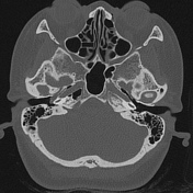

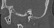
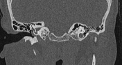
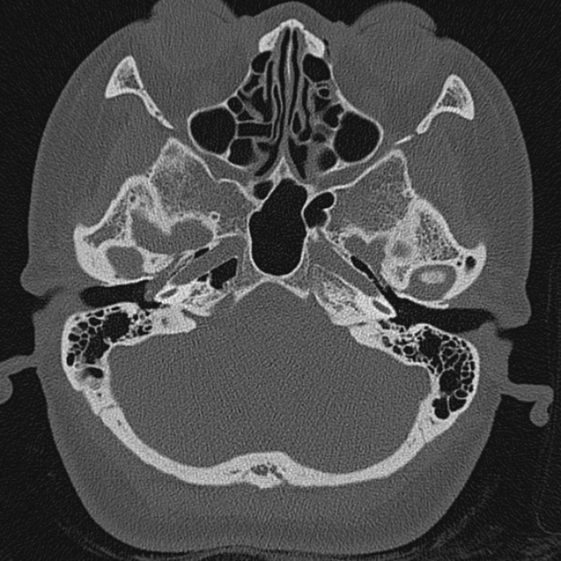
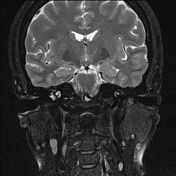
8mm well defined soft tissue mass lateral to the right cochlear promontary and posterior to the tympanic membrane.
No bony erosion. No extension into the inner ear.
Obstructive fluid partially opacifies the right mastoid air cells.
Ossicular chain normal.
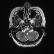
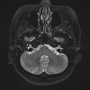
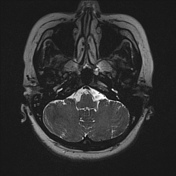

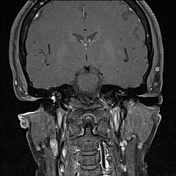

8 mm well-defined high T2, avidly enhancing soft tissue mass lateral to the right cochlear promontary and posterior to the tympanic membrane.
No internal cystic change or flow voids.
Fluid in the right mastoid air cells.
Reactive internal jugular chain lymph nodes.
Case Discussion
A nice example of a small tympanic paraganglioma (formerly known as glomus tympanicum), which correlates well with the patient demographics and clinical assessment.
The typical appearances are of a well defined mass with its base on the cochlear promontory, which avidly enhances on MRI. It is the most common tumor of the middle ear.


 Unable to process the form. Check for errors and try again.