
Presentation
Struck to right side of head. Left hemiparesis prior to loss of consciousness.
Patient Data
This case featured in our 2016 Trauma Radiology Course which is now available to view online.


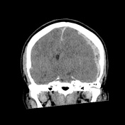
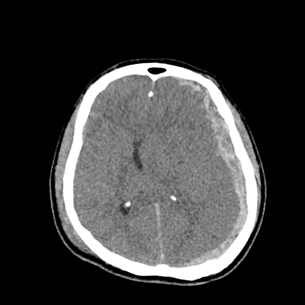
Right sided scalp hematoma and skull / petrous temporal bone fracture with contrecoup left cerebral convexity acute subdural hematoma producing substantial mass effect with midline shift and left uncal herniation. The uncal herniation is severe and well identified by observing the temporal horn of the left lateral ventricle descending below the tentorium. Small volume traumatic subarachnoid blood is also present and there are small right temporal intraparenchymal hemorrhages. In addition to the right petrous temporal fracture, there is hemorrhage within the sphenoid sinus indicating other skull base injuries.
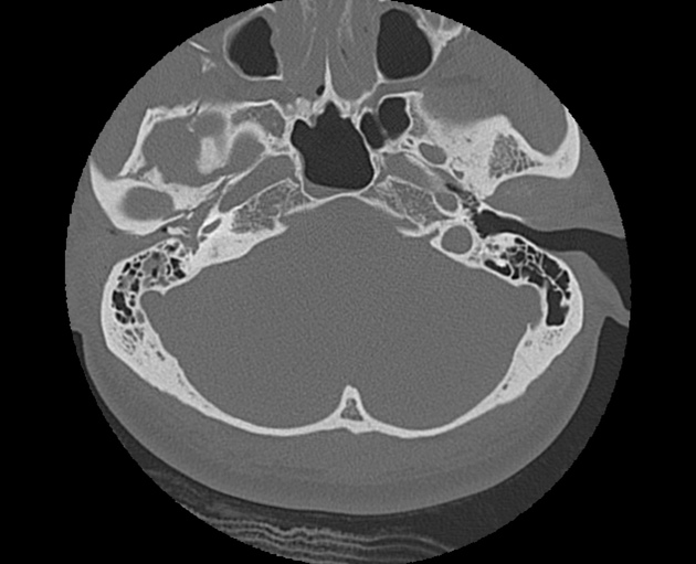
Predominately longitudinal fracture of the right petrous temporal bone with complete disruption of the ossicular chain. Normal anatomy is well demonstrated on the left side.
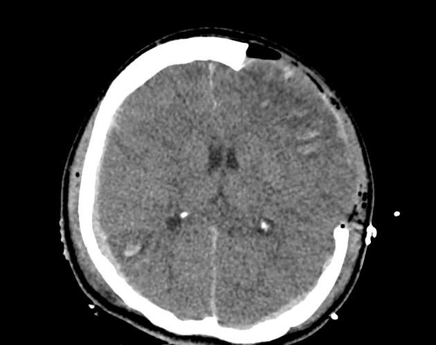
Post emergency left craniectomy and subdural evacuation. Mild herniation of brain out the craniectomy defect. While the midline shift and the uncal herniation have resolved, a greater size and number of intraparenchymal hemorrhages are now seen in addition to subdural blood on the tentorium and along the right cerebral convexity. It is common for this extension of intraparenchymal bleeding to occur once the tamponade effect of raised intracranial pressure is released. Importantly, there is now hypoattenuation and possible petechial hameorrhages evident within the right side of the midbrain. This has occurred to compression of this side of the midbrain against the tentorium when the contralateral uncal herniation was present (Kernohan notch) and accounts for the patient's left hemiparesis prior to losing consciousness (Kernohan phenomenon).
Case Discussion
This case teaches several important traumatic concepts;
- contrecoup subdural hematoma - i.e. on opposite side to head strike
- uncal herniation - look for the temporal horn of the lateral ventricle
- Kernohan phenomenon - contralateral midbrain compression due to uncal herniation
- increase in intraparenchymal hematoma size following decompressive craniectomy due to loss of tamponade effect from raised intracranial pressure
- association of longitudinal petrous temporal bone fracture with ossicular chain disruption


 Unable to process the form. Check for errors and try again.