Presentation
Left thigh pain and swelling associated with paraesthesia and heaviness for 3 months.
Patient Data
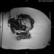

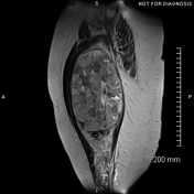

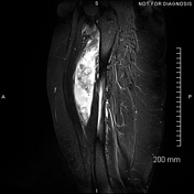

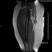

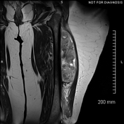

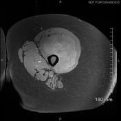

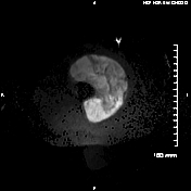

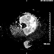

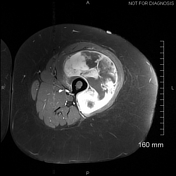

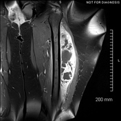

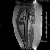

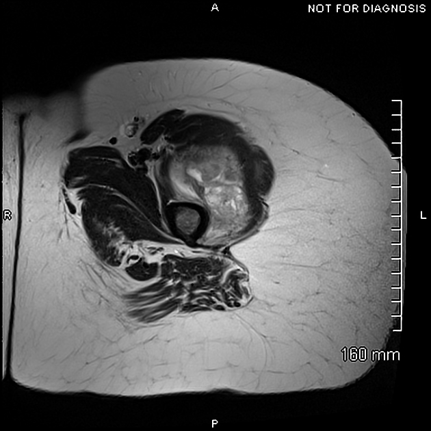
Large well-defined heterogeneous soft tissue mass lesion occupying most of the left vastus intermedius muscle. It measures about 17 x 11 x 10 cm and is isointense to muscles on T1 and mildly hyperintense on T2-weighted images. Mild diffusion restriction is noted on diffusion weighted images. Post-contrast images show extensive enhancement of the solid component and multiple non-enhancing necrotic areas. Extensive high T2 signal (oedema or infiltration?) is noted in the remaining portion of the muscle. Intact vastus lateralis, vastus medialis as well as rectus femoris muscle. No signs of invasion of the underlying femur. The neurovascular bundle (in the abductor canal) is intact.
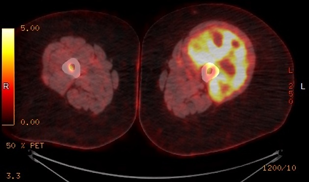
A heterogeneously hypermetabolic aggressive-looking mass involving the musculature of the lateral compartment of the left thigh. The lesion has a standardised uptake value of 23 and is in very close proximity to the femoral cortex.

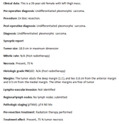
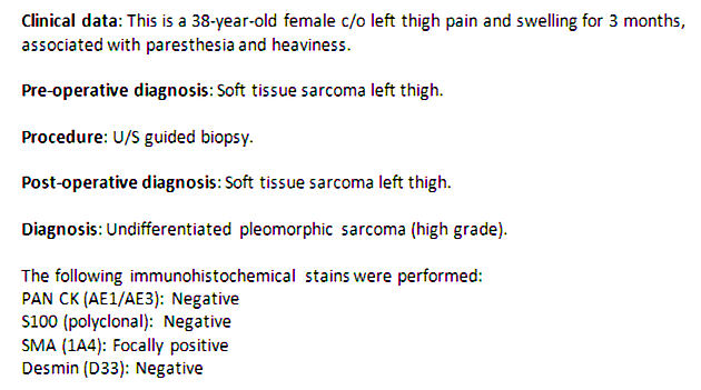
Histopathology showed undifferentiated pleomorphic sarcoma.
Case Discussion
Sizeable malignant-looking hypermetabolic mass involving the left vastus intermedius muscle. Imaging features are suggestive of soft tissue sarcoma like undifferentiated pleomorphic sarcoma (UPS), previously known as malignant fibrous histiocytoma (MFH), rhabdomyosarcoma or malignant peripheral nerve sheath tumour.


 Unable to process the form. Check for errors and try again.