
Presentation
Primary infertility and amenorrhea
Patient Data


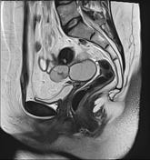
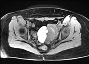
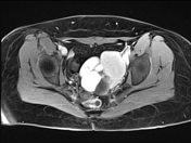
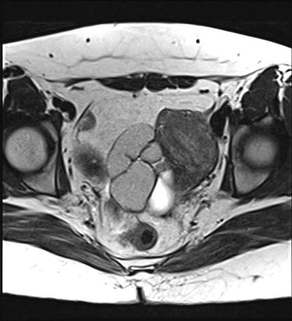
An abnormal contour of the uterus that is seen at the left side of the pelvis, appears curved and elongated giving a "banana-shaped" external contour. It appears of relatively reduced volume with a small endometrial cavity however, the myometrial zonal anatomy is preserved. The cervicovaginal canal appears small. No detectable communicating or non-communicating rudimentary horns.
Multilocular adnexal cystic lesion of tubular like configuration is seen related to the uterus elicits high signal in T1 with shading on T2 denoting hemorrhagic content.
Other multiple cysts are seen closely related to the uterus elicit fluid signal intensity likely left ovarian cysts.
The right ovary is seen more cranial at the right iliac fossa appears of average size and normal signal with few small follicles.
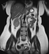
Left renal agenesis.
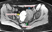

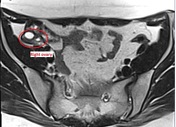

Annotated images clarifying the study findings.
Case Discussion
This case shows a unicornuate uterus with associated left renal agenesis "Müllerian duct anomaly class II". There is no rudimentary horn, consistent with "type b". There is associated hematosalpinx related to the drainage of the unicornuate uterus into the fallopian tube explaining primary amenorrhea. On initial clinical examination, there was a very small vagina, primarily concerning imperforate hymen or vaginal septum.


 Unable to process the form. Check for errors and try again.