
Presentation
Poor urinary stream and hematuria.
Patient Data
Age: 85 years
Gender: Male
From the case:
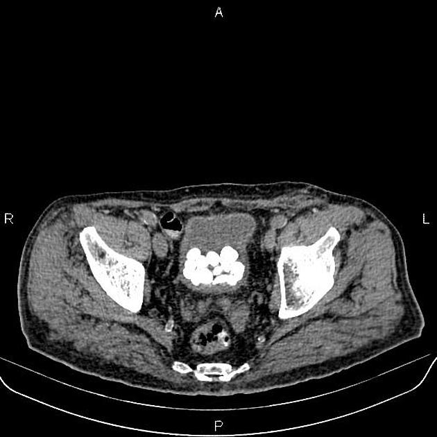
Urinary bladder stones


Download
Info
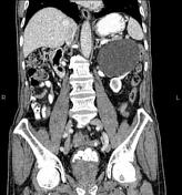
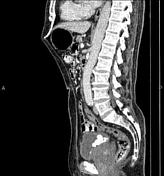
A 120×90 mm thin walled partially exophytic cyst is noted at left kidney without calcification or enhancing solid component. In addition, a few small simple cortical cysts are seen at kidneys less than 30 mm.
There is also a 14 mm (880 HU) stone is evident at left kidney.
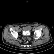
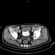
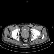
Diffuse but mild urinary bladder wall thickening is present. Numerous varying sized vesical stones are also observed less than 25 mm.
The prostate gland is markedly enlarged.
Degenerative changes as osteophytosis are seen at the lumbar spine.
Case Discussion
Bladder calculi occur either from migrated renal calculi or urinary stasis and may present with pain, infection, hematuria or may be asymptomatic.


 Unable to process the form. Check for errors and try again.