
Presentation
Menorrhagia with an irregular vaginal bleeding. History of a previous hysteroscopy for excision of a uterine mass.
Patient Data
Recent ultrasound study

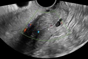
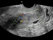
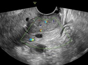
Ultrasound study shows:
- a bulky uterus with a cervical mass lesion is seen distending the cervical canal and extending through the internal os to the uterus with internal vascularity.
- multiple cervical nabothian cysts.
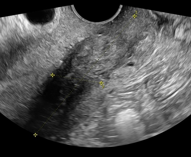
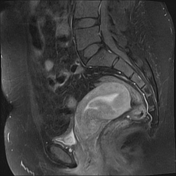
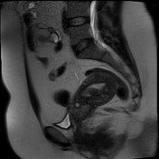
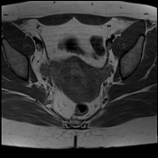
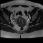
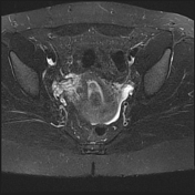
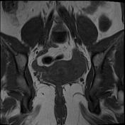
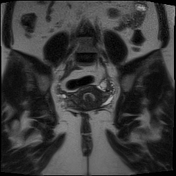
Recent MRI study
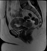

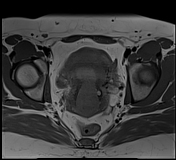
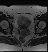
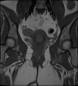
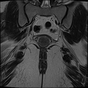

MRI study shows:
- a bulky anteverted uterus with a large cervical canal mass lesion measures 6x4x3.5 cm, seen extending through the internal os to the uterus, blending with its posterior wall and located in a sub endometrial location. The lesion elicits intermediate signal intensity in T1 and T2 with moderate enhancement in the post-contrast study
- multiple cervical nabothian cysts
The patient was subjected to hysteroscopic excision of the mass and confirmed with the histopathology to be a leiomyoma.
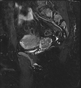
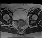
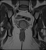
Old MRI study two years ago

MRI study shows a retroverted uterus with a posterior wall submucous leiomyoma (confirmed histopathologically).
Case Discussion
Anteversion is the most common uterine position in adults followed by retroversion that occurs in about 15-20% of women. Change of the uterus position from anteversion to retroversion may occur in certain conditions like bladder fullness or pregnancy however change from retroversion to anteversion is relatively uncommon 1.
Here is a case of recurrent submucosal uterine leiomyoma prolapsed into the cervical canal associated with uterine position change from retroversion to anteversion.
Ultrasound contribution by Dr.Shaimaa Hussien


 Unable to process the form. Check for errors and try again.