
Presentation
G1P0, 11 weeks by dates, with pelvic pain.
Patient Data
Age: 25 years
Gender: Female
From the case:
Uterine contraction in pregnancy


Download
Info

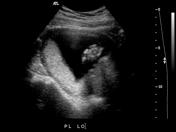
Left image: a focal retroplacental mass/bulge that is isoechoic to the uterus.
Right image: 1 week follow up with resolution of uterine contraction.
Case Discussion
Focal retroplacental mass/bulge that is isoechoic to the uterus. Differential considerations include:
Diagnosis
Follow-up was performed to confirm the diagnosis of uterine contraction. Note the resolution of the retroplacental bulge on follow-up imaging.
Doppler assessment is also useful (not shown here):
- hematoma- there is no Doppler flow
- uterine contraction- increased RI
- myoma - little Doppler flow


 Unable to process the form. Check for errors and try again.