
Presentation
55-year-old man with marked cognitive slowing and unsteady gait. Background of 10 year history of functional decline coinciding with onset of bipolar affective disorder. Frequent psychiatric inpatient admissions for mania in context of poor adherence with medications including sodium valproate. Medical history of severe ischemic heart disease, kidney failure and hypertension.
Patient Data


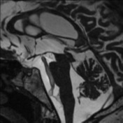
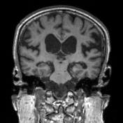

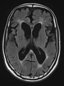
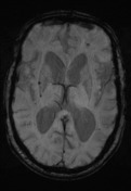

The lateral ventricles are dilated with extensive periventricular white matter T2 hyperintensity that is marked for age with associated diffuse white matter volume loss particularly in the frontal lobes. However there is relative crowding of the sulci at the vertex. Minimal upward bowing of the thinned corpus callosum. Although the walls of the 3rd ventricle are laterally bowed, the floor is not particularly depressed. The cerebral aqueduct is widely patent. Prominant aqueductal flow. Aqueductal stroke volume (noise corrected) is 107 microliters, elevated. Blooming artefact in the pons, basal ganglia and the left frontal lobe in keeping with foci of previous microhemorrhage with a distribution most in keeping with hypertensive bleeds.
Conclusion: Diffuse cerebral atrophy with a frontal predominance with evidence of chronic small vessel ischemic change. However, there are also findings suspicious for concomitant chronic communicating hydrocephalus/NPH, particularly when in the appropriate clinical context.
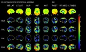
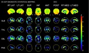
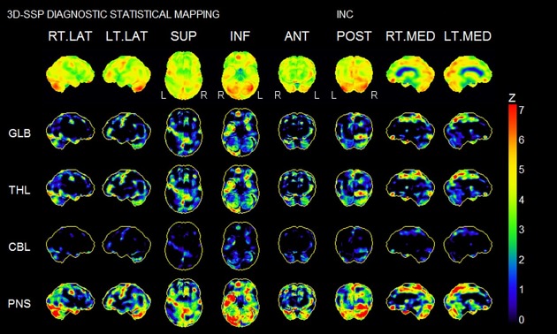
SPECT studies of the brain were performed with some reduction in image quality noted due to patient movement. The studies show focal hypoperfusion in the prefrontal lobes and in the opercular regions are superficial to the insula. Posterior cingulate and precuneus perfusion appears preserved without diagnostic features of Alzheimer's disease. Very small foci of posterior parietal hypoperfusion may be consistent with trauma or vascular disease. The anterior temporal lobes are well-perfused as are the basal ganglia and the cerebellum. Neurostat analysis also performed confirms the impression of free frontal and opercular hypoperfusion with the prominent enlarged lateral ventricles also being delineated. The studies are highly suspicious of the frontal variant of frontotemporal dementia. There may also be a vascular component of dementia present and ventriculomegaly is confirmed.
Neuropsychiatry Unit Cognitive Screening Tool
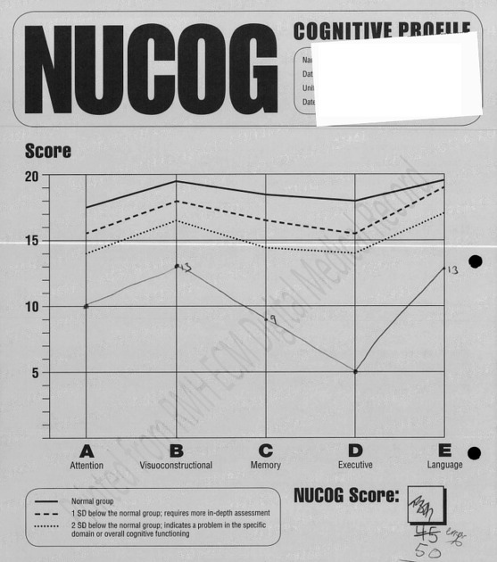
Cognitive testing was undertaken. NUCOG 50/100. Results across all domains were more than two standard deviations below normal.
Case Discussion
The key differentials in this case are normal pressure hydrocephalus (NPH) and sodium valproate encephalitis. The clinical and imaging findings however point towards a diagnosis of vascular dementia. The MRI shows a lot of white matter change. The ventricles are widened. Of note, and against the diagnosis of NPH is that he has a patent cerebral aqueduct and his callosal angle measured in the coronal plane is within the lower limits of normal.


 Unable to process the form. Check for errors and try again.