
Presentation
The patient was reviewed for abdominal discomfort at both flanks, pain during passing urine, and difficulty to pass urine. 2-5 RBC S are noted in the urine analysis.
Patient Data


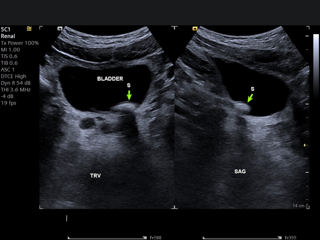
The ultrasound transverse and sagittal sections to the left lower ureter shows a large ureteric stone measuring about 17x10 mm in the left vesicoureteric junction.
Minimal renal fulness is noted in the left kidney.
Case Discussion
The hydronephrosis is not always seen in the ureteric stone, sometimes the stone doesn't cause renal dilation.
There is no sure relationship between the ureteric stone size and the hydronephrosis degree, sometime small stones may cause mild to moderate hydronephrosis, while a big one may cause minimal or mild hydronephrosis or just slight fullness in the renal pelvis which may become clear if patient drink water.


 Unable to process the form. Check for errors and try again.