Presentation
Headache and decreased vision.
Patient Data
![](https://prod-images-static.radiopaedia.org/images/14401584/6d00542096dfd1f470588a8aed5a0f_thumb.jpeg)
![This study is a stack](/packs/stack-YQKLCKBI.gif)
![](https://prod-images-static.radiopaedia.org/images/14397907/7877f6420de7f748a29e3cc2351c2e_thumb.jpeg)
![This study is a stack](/packs/stack-YQKLCKBI.gif)
![](https://prod-images-static.radiopaedia.org/images/14401291/3d60963e2ea001d9e73b19eb5f4cee_thumb.jpeg)
![This study is a stack](/packs/stack-YQKLCKBI.gif)
![](https://prod-images-static.radiopaedia.org/images/14402802/9c9fea7bfcaf7c1cd530552bf2e534_thumb.jpeg)
![This study is a stack](/packs/stack-YQKLCKBI.gif)
![](https://prod-images-static.radiopaedia.org/images/14400670/497030e8c9fae407981d241d74eb49_thumb.jpeg)
![This study is a stack](/packs/stack-YQKLCKBI.gif)
![](https://prod-images-static.radiopaedia.org/images/14403351/5385e3ec9282c906003ad1dd0f3118_thumb.jpeg)
![This study is a stack](/packs/stack-YQKLCKBI.gif)
![](https://prod-images-static.radiopaedia.org/images/14405012/5b989a51817a279383e133b82199c7_thumb.jpeg)
![This study is a stack](/packs/stack-YQKLCKBI.gif)
![](https://prod-images-static.radiopaedia.org/images/14400017/359297015f4812c90792a915f2c463_thumb.jpeg)
![This study is a stack](/packs/stack-YQKLCKBI.gif)
![](https://prod-images-static.radiopaedia.org/images/14404117/73a88dece4d2f6ad1e6d375f9b6a03_thumb.jpeg)
![This study is a stack](/packs/stack-YQKLCKBI.gif)
![](https://prod-images-static.radiopaedia.org/images/14404859/78d58855d09ff8931a341db3bcb1f1_thumb.jpeg)
![This study is a stack](/packs/stack-YQKLCKBI.gif)
![](https://prod-images-static.radiopaedia.org/images/14403514/d75110b3f6caab3d8e55e27547a7ee_thumb.jpeg)
![This study is a stack](/packs/stack-YQKLCKBI.gif)
![](https://prod-images-static.radiopaedia.org/images/14404280/842185a368e84701fd5920156eda28_thumb.jpeg)
![This study is a stack](/packs/stack-YQKLCKBI.gif)
![](https://prod-images-static.radiopaedia.org/images/14403808/74cf7c32b9db1c3748cde1dd20303e_thumb.jpeg)
![This study is a stack](/packs/stack-YQKLCKBI.gif)
![](https://prod-images-static.radiopaedia.org/images/14404561/0bee14ae1adee09b452ebb09f76ce7_thumb.jpeg)
![This study is a stack](/packs/stack-YQKLCKBI.gif)
![](https://prod-images-static.radiopaedia.org/images/14400373/7a48c7aefd2d7f97a09d5f60dbccb8_thumb.jpeg)
![This study is a stack](/packs/stack-YQKLCKBI.gif)
![](https://prod-images-static.radiopaedia.org/images/14403093/8feb8ec356a285312801d51cff11d6_thumb.jpeg)
![This study is a stack](/packs/stack-YQKLCKBI.gif)
![](https://prod-images-static.radiopaedia.org/images/14400964/08d946c8c7ab5130d570d7862aed85_thumb.jpeg)
![This study is a stack](/packs/stack-YQKLCKBI.gif)
![](https://prod-images-static.radiopaedia.org/images/14402415/0b9023fcc45f912e2d0a3d74b7f7c8_thumb.jpeg)
![This study is a stack](/packs/stack-YQKLCKBI.gif)
![](https://prod-images-static.radiopaedia.org/images/14401584/6d00542096dfd1f470588a8aed5a0f_big_gallery.jpeg)
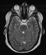
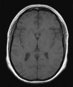
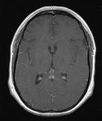
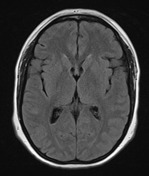
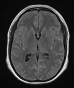
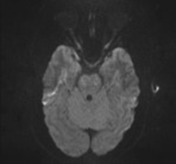
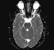
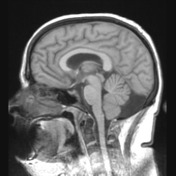
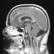
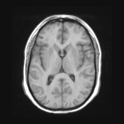
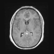
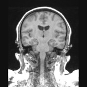
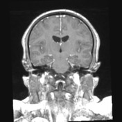
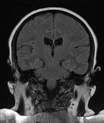
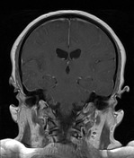
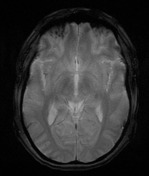
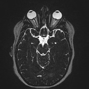
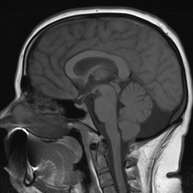
MRI of the brain demonstrates prominent sulcal high T2 signal on post Gad FLAIR and much less florid leptomeningeal enhancement. Prominent papilledema is visible, best seen on thin T2. Otherwise the study is unremarkable.
Case Discussion
The patient went on to have a lumbar puncture.
Cerebrospinal fluid
- CSF Protein 2.99 g/L (Normal <0.45)
- CSF Glucose 6.1H mmol/L (Normal 2.2 - 3.9)
- CELL COUNT: x10^6/L
- Erythrocytes 294
- Polymorphs 35
- Lymphocytes 180
BACTERIAL CULTURE No growth after 6 days
FUNGAL CULTURE : No growth after 14 days
CRYPTOCOCCAL SEROLOGY (CSF and Serum): Cryptococcal Ag: NOT Detected
-
MYCOBACTERIUM
DNA Amplification Assay for M.tuberculosis Complex(PCR): Negative
CULTURE SCREEN: MGIT bottle: Negative
MACROSCOPIC APPEARANCE: CSF Total volume: 10.0 ml(s), Slightly cloudy
MICROSCOPIC DESCRIPTION: The smears contain abundant lymphocytes with some monocytes. The lymphoid cells show no nuclear atypia. No increased numbers of neutrophils or eosinophils are seen. No organisms are present. No malignant cells are identified. Auramine-Rhodamine Stain: No Acid Fast Bacilli Detected (A negative acid-fast smear result does not exclude the presence of mycobacterium species.)
FINAL DIAGNOSIS: Chronic inflammation. No evidence of malignancy.
Discussion
Features are consistent with viral meningitis. The patient gradually recovered with only supportive measures.
Post contrast FLAIR is very helpful in identifying any condition with leptomeningeal vessel leakiness. It is therefore helpful in meningitis and leptomeningeal carcinomatosis.