
Presentation
Left loin to groin pain. Increasing urinary frequency.
Patient Data
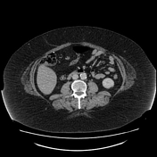

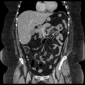

There is a 4 mm calculus within the urinary bladder within a subtle rounded cystic structure protruding from the left VUJ. Hypo-enhancement of the left renal upper pole and mild prominence of the pelvicalyceal system. Duplex collecting system with no dilatation of the upper or lower pole moieties. The right renal tract is unremarkable. No significant bladder wall thickening or pericystic fat stranding.
No intraperitoneal free fluid or free gas. VP shunt tubing was noted in the upper right abdomen. No associated intra-abdominal collection.
The liver demonstrates homogeneous reduced attenuation in keeping with steatosis. No focal liver lesions. The gallbladder, pancreas, cysts, spleen, and adrenal glands appear unremarkable. Previous gastric surgery and small hiatus hernia. Small and large bowel are unremarkable.
Impression
Left bladder calculus within a ureterocoele with features of a left duplex system. Left renal upper pole hypo-enhancement could be due to recent mild obstruction secondary to the ureterocoele calculus or superimpose
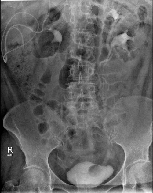
Opacification of the urinary collecting systems and bladder secondary to recent CT contrast administration.
A duplex collecting system is demonstrated on the left, with a round 2.7 cm filling defect at the level of the left VUJ representing a ureterocoele. The upper moiety does not appear obstructed.
The known small calculus is not appreciated.
Case Discussion
The patient had passed the small calculus with complete symptom relief in the hour between the CT and X-ray. The patient was afebrile and urine clear of leucocytes and nitrites.


 Unable to process the form. Check for errors and try again.