
Presentation
Left parotid lump.
Patient Data


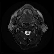
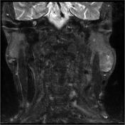
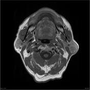
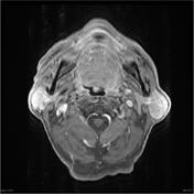
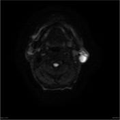
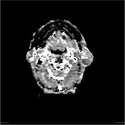
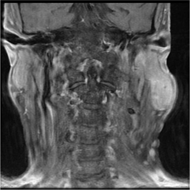
Rounded mass seen in the retromandibular left parotid tail measuring 1.5 by 1.8 centimeters in axial dimensions and 2.6 centimeters superoinferiorly. There is heterogeneous internal signal including small areas of T1 hyperintensity, and heterogeneous enhancement. There are smaller adjacent nodules in the parotid, measuring 11.5 x 6.2 mm in the left parotid tail and 11.3 x 5.3 mm superiorly in the left parotid. These may be small lymph nodes. No abnormal perineural enhancement.
No right parotid lesions are seen. The right submandibular gland appears atrophic (not shown)
Encephalomalacia is seen at the left temporal pole suggestive of prior trauma.
Tiny T2 hyperintense nodule subpleural in the left lobe of the thyroid. Mucosal thickening in the left maxillary sinus.
Conclusion:
Findings compatible with a left parotid Warthin's tumor. Small adjacent intraparotid nodules likely lymph nodes.
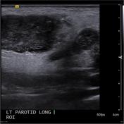
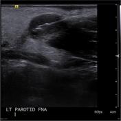
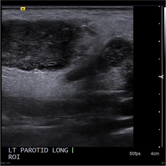
Within the left parotid gland there is a large 3.0 x 1.5 x 1.3 cm well- defined hypoechoic lesion. A smaller adjacent similar lesion. The larger lesion demonstrates mild heterogeneity and internal vascularity. No posterior acoustic shadowing. A core biopsy of this lesion was performed.
Technique:
Informed consent obtained. 1% lignocaine used for local anesthesia. Under ultrasound guidance a core biopsy was performed with a 20g QuickCore biopsy system. Sample was sent for analysis in formalin. No immediate post procedure complications.
Histology
MICROSCOPIC DESCRIPTION: The sections show a fragmented core biopsy which consists of prominent oncocytic cells with associated benign lymphoid tissue. There is no evidence of malignancy seen.
DIAGNOSIS: Left parotid lesion core biopsy: Features in keeping with a Warthin's tumor.
Case Discussion
Key learning points:
1. A heterogenous small parotid lesion <2cm is unlikely to be a pleomorphic adenoma.
2. Warthin tumors are most common in elderly male smokers and can be bilateral.


 Unable to process the form. Check for errors and try again.