
Presentation
Three weeks of vertigo.
Patient Data
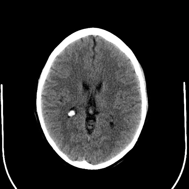
A lobulated mass is interposed between the medulla and the clivus.
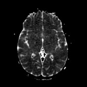
This demonstrates moderately increased density relative to the adjacent brain. The cerebrum is unremarkable.
No cerebellar abnormality is identified.
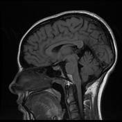

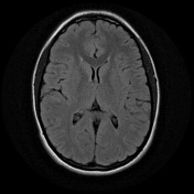
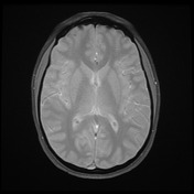
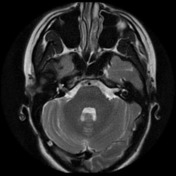
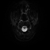
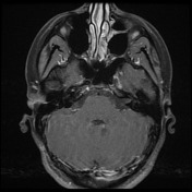
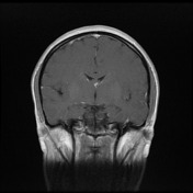
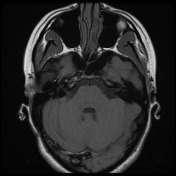

Evidence of prior right occipital craniotomy and stable appearance of multilobulated T1 hyperintense T2 hypointense non-enhancing pre-pontomedullary extra-axial lesion. The lesion lies centrally and to the right of the pre pontine and perimedullary cistern insinuating around the contour of the pons and the medulla displacing the basilar artery anteriorly with minor associated vessel narrowing. No significant mass effect on the adjacent brainstem. Patchy diffusion restriction. No contrast enhancement.
The patient proceeded to craniotomy and resection.
Histopathology
MICROSCOPIC DESCRIPTION: The section shows an extremely small fragment of laminated keratin consistent with the contents of either an epidermoid or dermoid cyst. No epithelial lining is included.
DIAGNOSIS: Prepontine lesion: Small fragment of keratin consistent with contents of either an epidermoid or dermoid cyst.
Case Discussion
This "white epidermoid" is so-called because of the intrinsic high T1 signal, which is atypical for epidermoid cysts.


 Unable to process the form. Check for errors and try again.