
Presentation
Abdominal pain and left side fullness on physical exam.
Patient Data
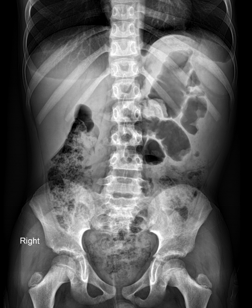
A significant mass effect is seen in the left abdominal cavity that displaces the spleen superiorly and colon loops medially.
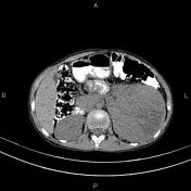

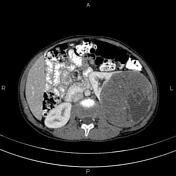
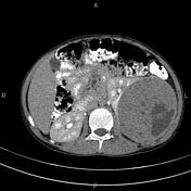
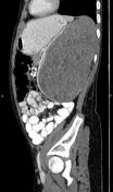


A large hetero-dense well-defined mass with few internal calcification foci and claw sign is seen in the left kidney. No frank fat density is noted within. After contrast media injection the mass shows heterogeneous enhancement with areas of internal cystic/necrotic changes.
There is no sign of local invasion, vascular extension, or regional lymphadenopathy.
Case Discussion
Typical appearance of Wilms tumor in a 12-year-old boy.
The patient underwent a left nephrectomy and histopathology evaluation was confirmed.
On CT and MR images, Wilms tumors are heterogeneous density masses with infrequent areas of calcification and rarely fat density. In contrast media administration, patchy heterogenous enhancement with areas of cystic/necrotic changes could be seen. Up to 20% of cases have lung metastases at the time of diagnosis. The Claw sign can be helpful for the renal origin of the mass.


 Unable to process the form. Check for errors and try again.