
Patient Data


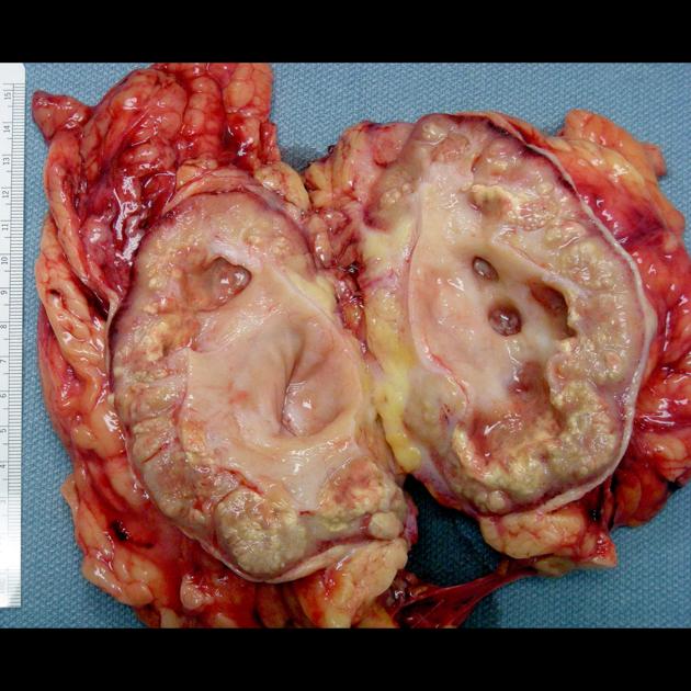
The specimen is received in OR and consistent with a resected kidney with abundant perirenal adipose tissue. The kidney is bivalved during intraoperative surgical pathology gross consultation. The dilated renal pelvis is filled with a large amount of pus material. The pus is sampled for culture. After washing the specimen, the walls of dilated calyces and renal pelvis are thickened with multiple yellow nodules. The renal cortex is largely atrophic.
Image courtesy of Jian-Hua Qiao, MD, FCAP, Los Angeles, CA, USA. Please see case description page for licence and original file information.


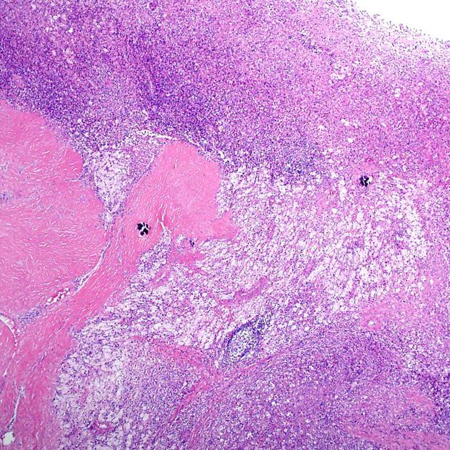
Sections through the yellow nodules show replacement of renal parenchyma with foamy histiocytes, occasional multinucleated giant cells, and inflammatory cells. The inflammatory cells are composed of lymphocytes, plasma cells, and neutrophils. Lymphoid aggregates are also noted. A few renal tubules are filled with cellular debris and possible bacterial clumps. Features are those of xanthogranulomatous pyelonephritis.
Image courtesy of Jian-Hua Qiao, MD, FCAP, Los Angeles, CA, USA.. Please see case description page for licence and original file information.
Case Discussion
This is a case of xanthogranulomatous pyelonephritis in a 35-year-old female.
Author: Jian-Hua Qiao, MD, FCAP, Los Angeles, CA, USA.
Original file: http://www.pathxchange.org/case/9372 (dead link)
Modifications: square crop
License: Image reproduced here with explicit permission of the author. All rights reserved by the author.
Permission ID: P-00014


 Unable to process the form. Check for errors and try again.