
Zygomaticomaxillary complex fracture, orbital blow-out fracture and a pituitary macroadenoma
Presentation
Road traffic accident
Patient Data

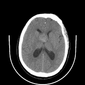

A relatively well-circumscribed midline homogeneously hyperdense soft tissue density lesion of ~44 to 54HU is noted occupying sellar and suprasellar region showing a mass effect on foramen of Monro with resultant moderate dilatation of bilateral lateral ventricles (right atrium ~1.8 cm, left atrium~2 cm). The lesion measures about (4 x 3.2 x 3 cm), in craniocaudal, transverse, anteroposterior dimensions respectively.
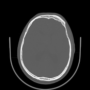
Comminuted fracture of the right zygomatic arch is noted with minimal displacement.
Fracture at posterolateral wall and floor of right orbit with minimal displacement is noted.
Mildly comminuted fracture with depression is noted at the medial wall of right orbit showing mild herniation of orbital fat.
Minimal right pneumoorbit is noted.
Undisplaced fracture lines are seen in the right maxillary sinus.
Fracture at the right lateral pterygoid plate is noted.
Soft tissue contusion with hematoma formation seen at the right frontal, periorbital and maxillary region.
Case Discussion
This case demonstrates the typical CT features of a pituitary macroadenoma, right zygomaticomaxillary complex fracture in combination with a blow-out type fracture of the right medial orbital wall.


 Unable to process the form. Check for errors and try again.