
1/ Asking “How old are you?” can be dicey—both in real life & on MRI! Do you know how to tell the age of blood on MRI?
Here’s a thread on how to date blood on MRI so that the next time you see a hemorrhage, your guess on when it happened will always be in the right vein!

2/ If you ask someone how to date blood on MRI, they’ll spit out a crazy mnemonic about babies that tells you what signal blood should be on T1 & T2 imaging by age. But mnemonics are crutch—they help you memorize, but not understand. If you understand, you don’t need to memorize
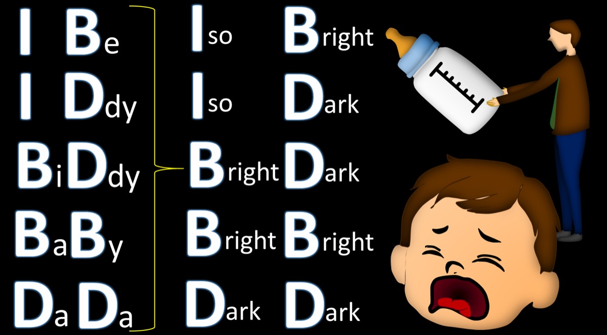
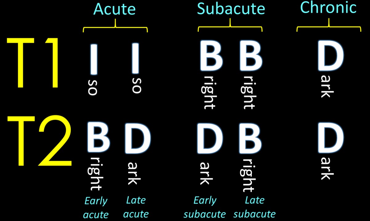
3/ If you look at the mnemonic, you will notice one thing—the T1 signal is all you need to tell if blood is acute, subacute or chronic.
T2 signal will tell if it is early or late in each of those time periods—but that type of detail isn’t needed in real life
So let’s look at T1
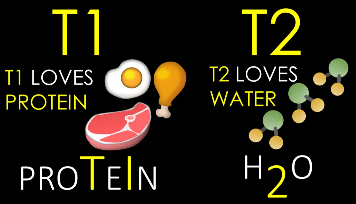
4/ To understand how blood looks on MRI at different ages, you need 2 basic MRI rules:
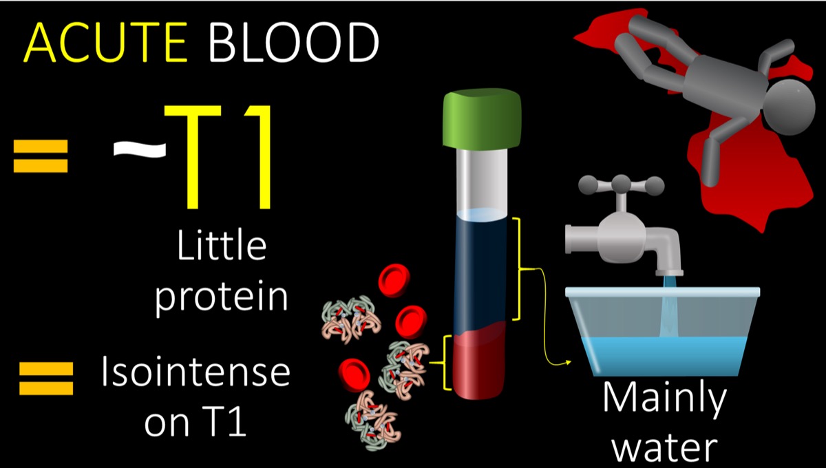
Remember T1 loves protein. More protein = brighter on T1. I remember this bc T1 looks like the T & I in protein
T2 loves water. Fluid is bright on T2. This is easy bc there’s a 2 in H2O.
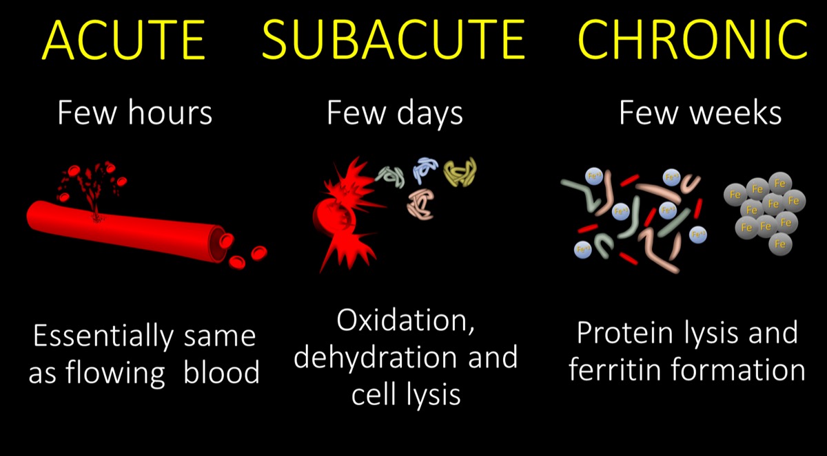
5/ Acute blood is in the first hours. It’s basically blood that has just poured out of the artery. If you think about how acute bleeding looks in real life, you know the properties of acute blood—it’s basically water w/a little protein to gives it the red color & thickness
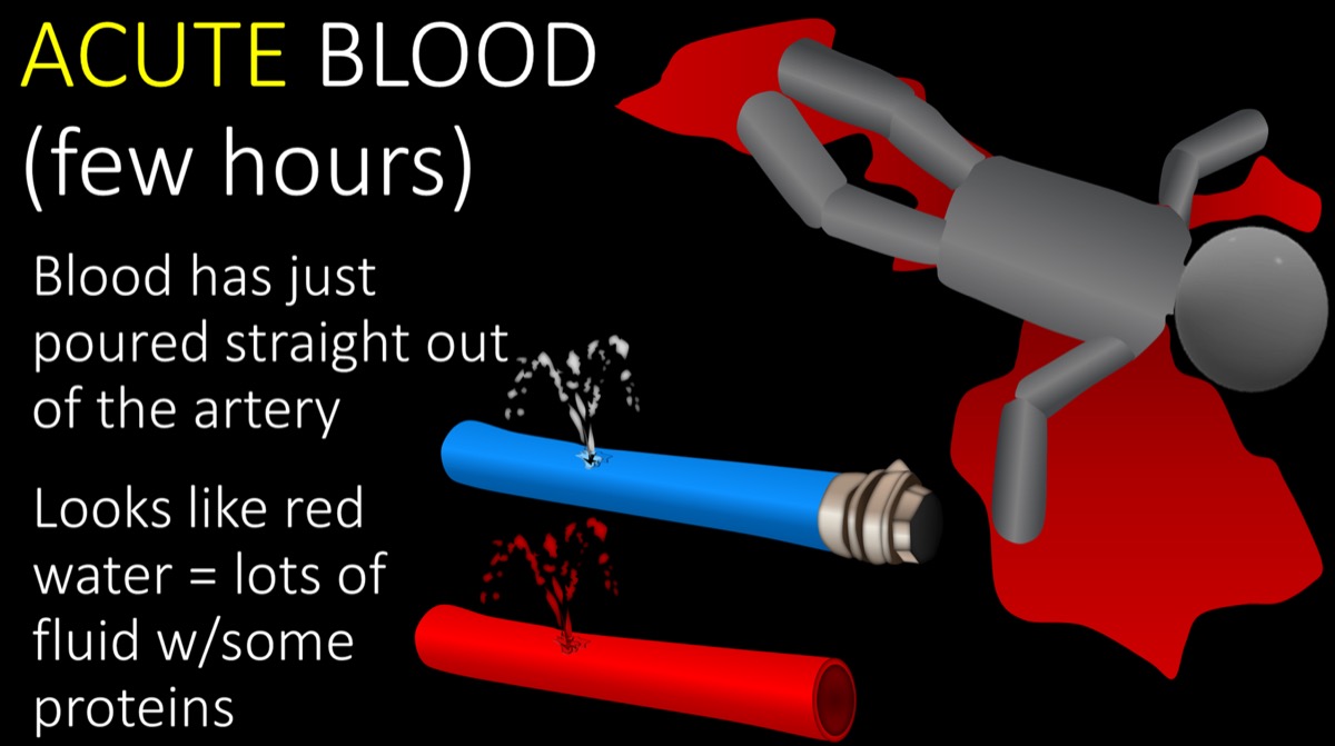
6/ How does T1 feel about acute blood? Well, acute blood is a lot of water w/a little protein So you will get some love from T1 for the little protein. But it won’t be super bright because the protein content isn’t that high—it’s diluted. So it acute blood is isointense on T1.
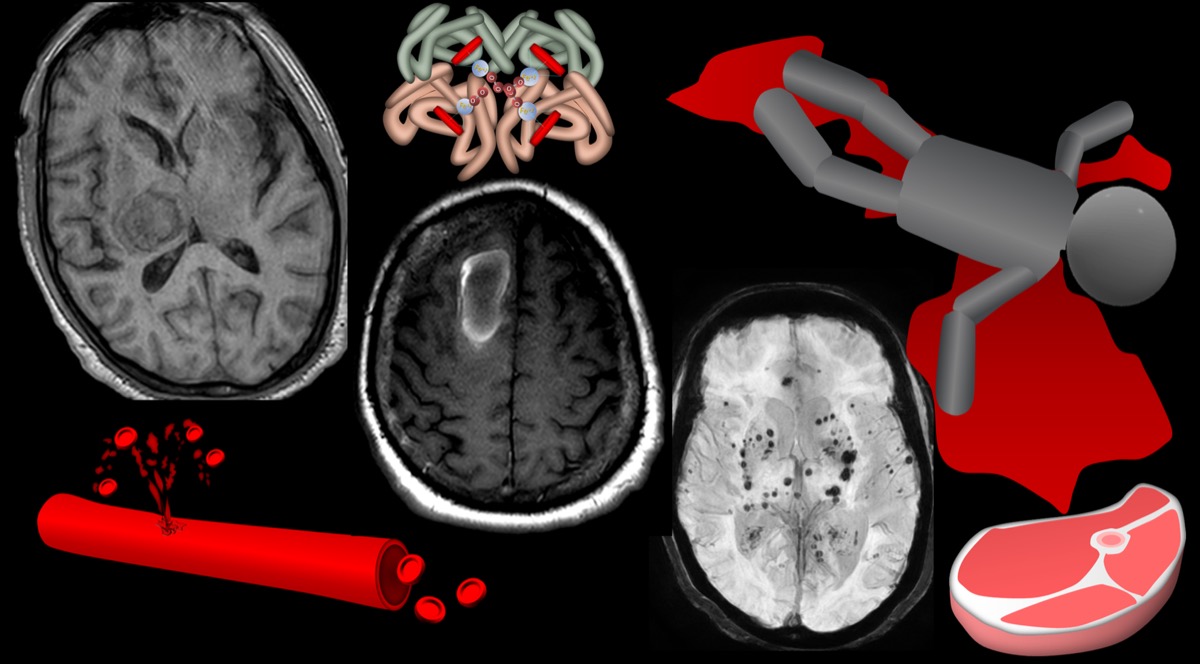
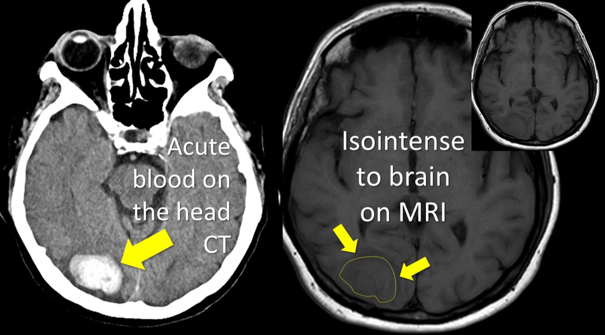
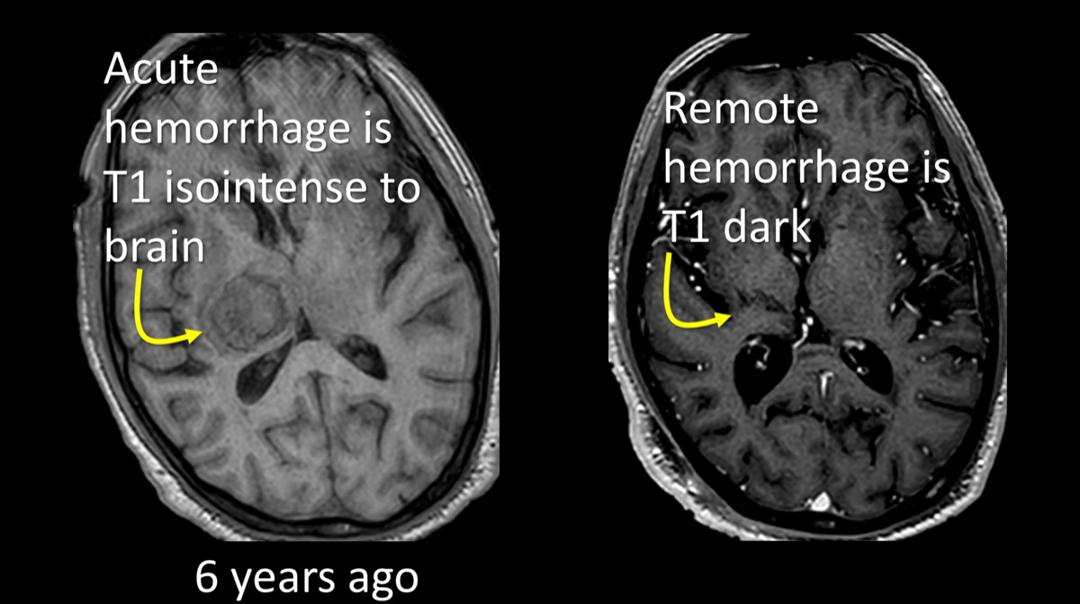
7/ Here’s an example of acute blood on T1. The hematoma is very dense on CT, consistent w/acute timing. On T1, it isointense to brain parenchyma. It’s not bright because protein content is relatively low. But it isn’t dark either, because blood has some protein that gives it some signal
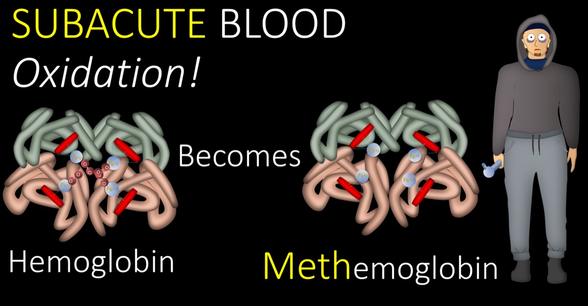
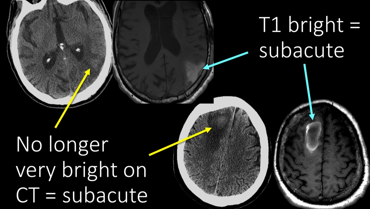
8/ After a few days, you get subacute blood. In the subacute period, blood gets oxidized. It’s like what happens to an apple when you leave it out, or why a steak turns dark when it’s left out. Subacute blood is oxidized blood that has been left out for a few days like a steak.
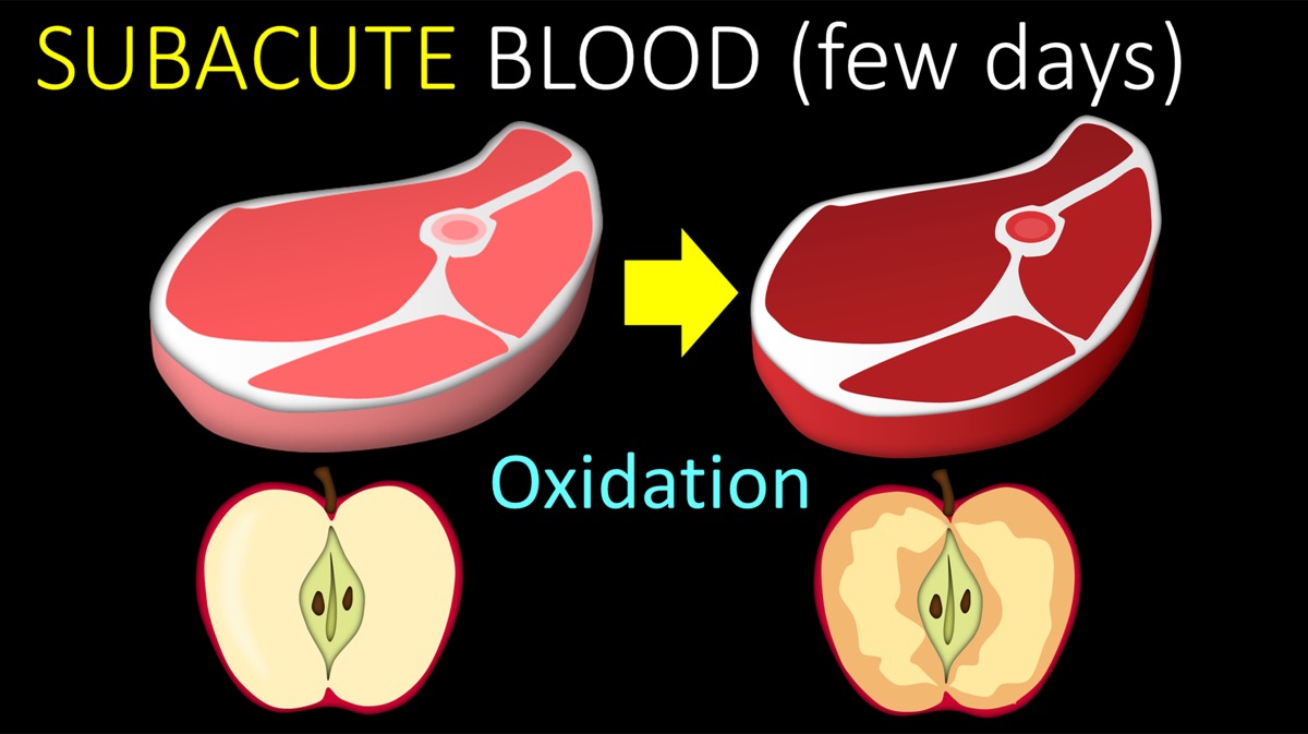
9/ When blood gets oxidized in the subacute period, hemoglobin becomes methemoglobin. This change in hemoglobin marks the transition from acute to subacute blood.
10/ Subacute period is like aging a steak. Cells will begin to lyse & water content will be lost. This is exactly what happens w/a steak. It’s why we age a steak—the broken down proteins & lower water content lead to a more tender & flavourful steak
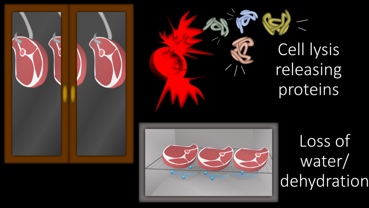
11/ Both of these processes—letting proteins out of cells & decreasing water content—will increase the density of protein. More protein means higher T1 signal. This contributes to giving subacute hematomas a very bright signal on T1.
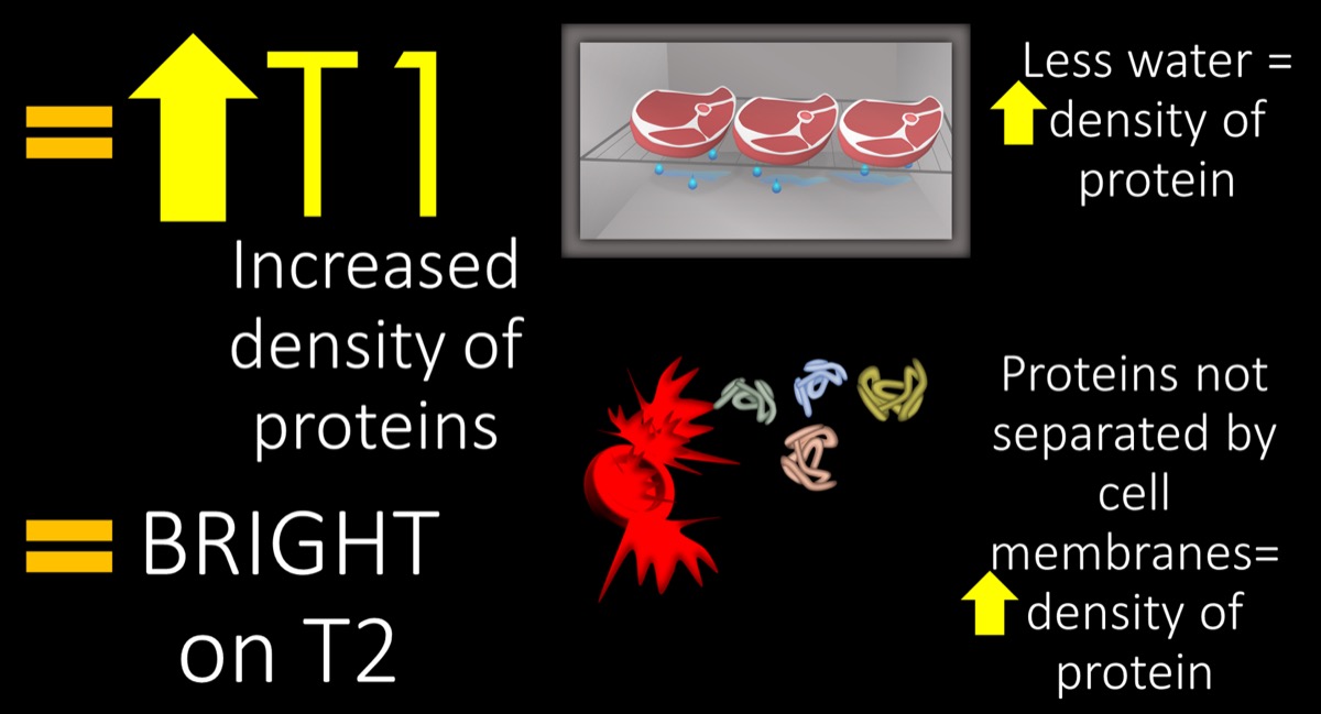
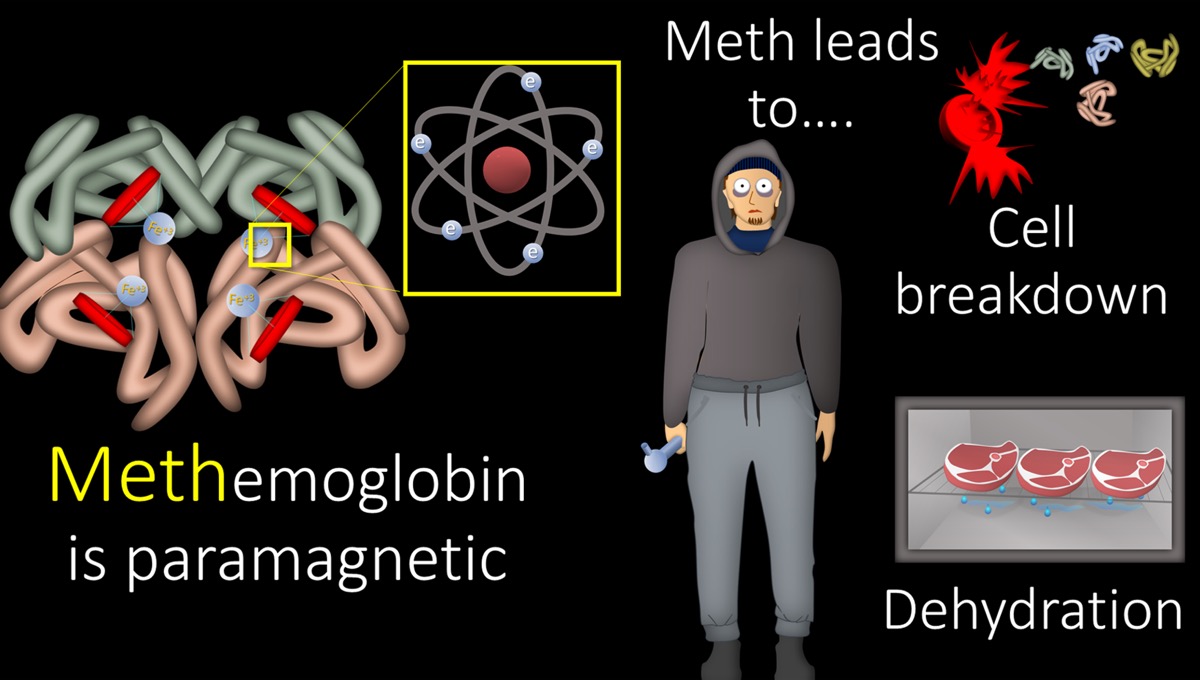
12/ Although more protein from the aging process leads to high T1, high T1 comes also from new electrons from the oxidation to methemoglobin. I just remember that using Meth is basically a way to age humans like dry aging a steak, so Meth(emoglobin) will lead to increased T1.
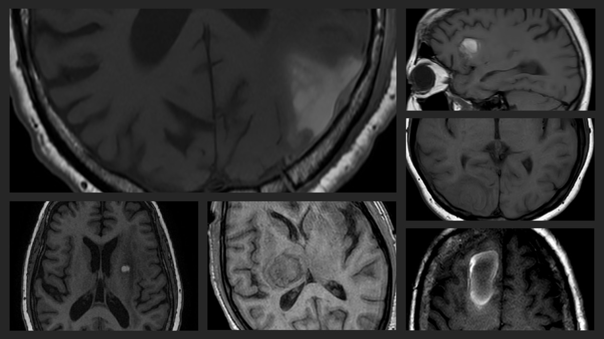
13/ Here are examples of subacute blood on MRI. You can see these hematomas are only subtly bright on CT now, as their acute clots have begun to be broken down. On MRI, these have increased T1 signal related to the increased protein and increased Meth.
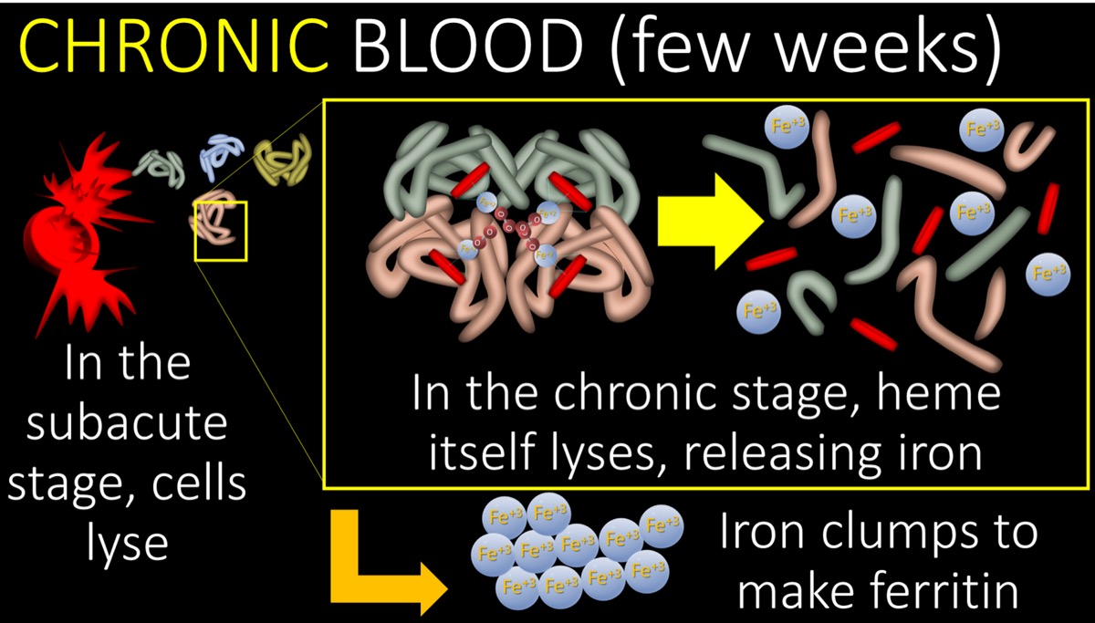
14/ Chronic blood is after a few weeks. In the subacute phase, cells lyse. In the chronic phase, the proteins themselves lyse, including heme. This releases the iron from the heme. The iron molecules from the broken heme start to clump together to make ferritin or hemosiderin.
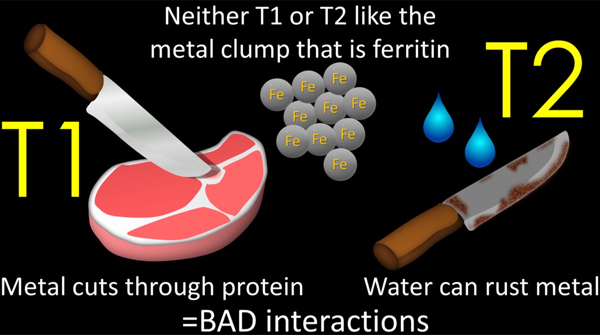
15/ So neither T1 or T2 sequences like metals like ferritin/hemosiderin. You can remember this because metal doesn’t mix well with protein (cuts right through it) or water (together water and iron make rust).
16/ Because neither T1 or T2 like ferritin/hemosiderin, you will end up getting a dark signal on both sequences in the chronic phase. In fact, no sequence really likes hemosiderin, and it will be dark on all sequences.
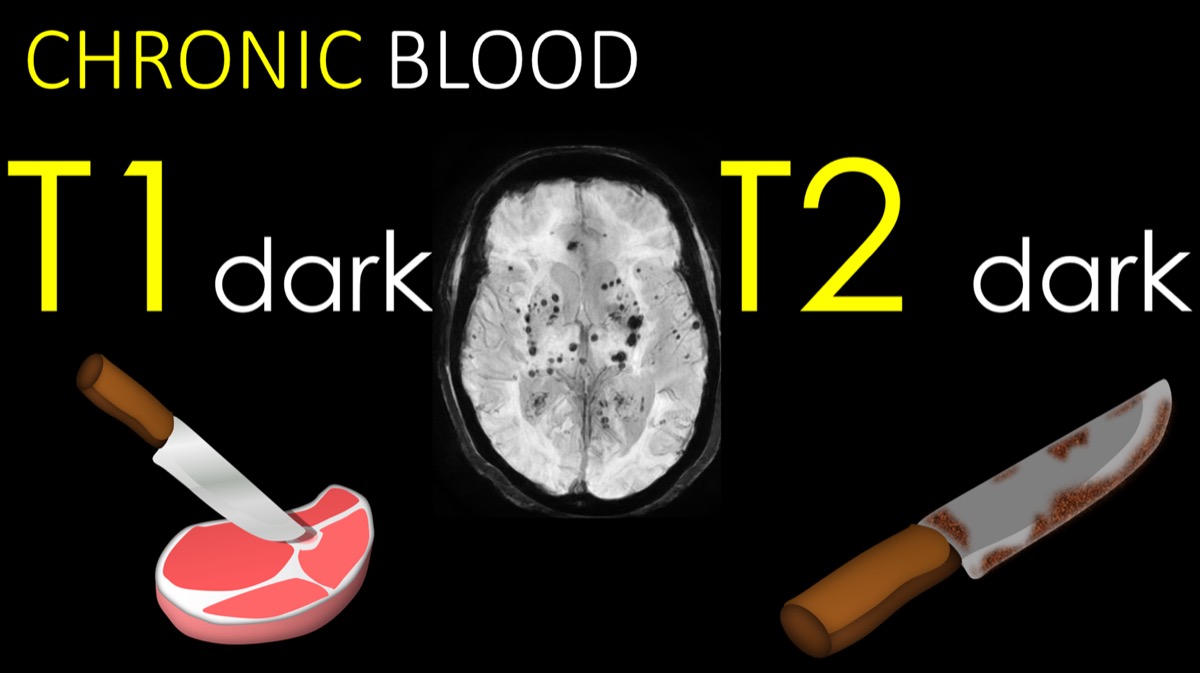
17/ Here’s an example of a chronic hematoma. Six years ago, it looked like an acute hematoma—isointense on T1 (bc there are some proteins in the fluid that is acute blood). Now it is dark on T1, bc everyone hates hemosiderin.
18/ So remember: acute is a few hours, subacute is a few days, & chronic is a few weeks. Acute blood is like flowing blood but outside the vessel Subacute blood has started oxidation & cell lysis. Chronic blood has broken down everything so that even iron is out on its own
19/ Knowing what acute, subacute, and chronic blood consist of can help you to remember your T1 signal:
- Acute is fluid w/little protein = iso
- Subacute has lots of protein from cell lysis & water loss & methemoglobin = bright
- Chronic is filled w/iron no one likes = dark

20/ T1 feels about blood like you feel about a good steak:
Acute is raw meat—has potential, but you won’t eat it yet = iso
Subacute has freed all the proteins for good taste, you want to dig in = bright
Chronic has broken down too much & is rotten—no thanks = dark.

21/ Of course, there are subtleties to this based on oxygen tension, blood flow to the region, hematocrit, etc.
But as a rule of thumb, think of blood on MRI like you would a good steak—bon appetite!


 Unable to process the form. Check for errors and try again.