Accessory ossicles
Disclosures
- updated 5 Sep 2023:
Nothing to disclose
Updates to Article Attributes
Body
was changed:
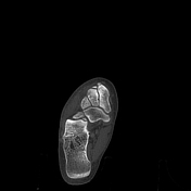
Accessory ossicles are secondary ossification centres that remain separate from the adjacent bone. They are usually round or ovoid in shape, occur in typical locations and have well-defined smooth cortical margins on all sides.
In most cases, they are congenital in origin, although they may occur as a result of trauma or local degenerative disease 2.
The significance of accessory ossicles is their potential to mimic avulsion fractures.
-
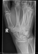
accessory ossicles of the wrist
trapezium secondarium
epilunate
-
accessory ossicles of the spine
-
accessory ossicles of the hip
-
accessory ossicles of the knee
-<li><p><a href="/articles/os-sesamoideum-tricipitale">os sesamoideum tricipitale</a> </p></li>- +<li><p><a href="/articles/os-sesamoideum-tricipitale">os sesamoideum tricipitale</a> </p></li>
-<p><a href="/articles/accessory-ossicles-of-the-wrist" title="accessory ossicles of the wrist">accessory ossicles of the wrist</a></p>- +<p><a href="/articles/accessory-ossicles-of-the-wrist" title="accessory ossicles of the wrist">accessory ossicles of the wrist</a></p>
-<li><p><a href="/articles/persistent-ossiculum-terminale">persistent ossiculum terminale</a> - Bergmann ossicle</p></li>- +<li><p><a href="/articles/persistent-ossiculum-terminale">persistent ossiculum terminale</a> - Bergmann ossicle</p></li>
-<p><a href="/articles/accessory-ossicles-of-the-foot" title="accessory ossicles of the foot">accessory ossicles of the foot</a></p>- +<p><a href="/articles/accessory-ossicles-of-the-foot" title="accessory ossicles of the foot">accessory ossicles of the foot</a></p>
Images Changes:
Image 1 Diagram ( create )

Caption
was added:
Figure 1: the foot
Position
was set to
1.
Image 2 Annotated image ( update )
Caption
was changed:
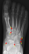
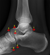
Figure 12: accessory ossicles of the foot
Position
changed from 1 to 2.
Image 3 Annotated image ( update )
Caption
was changed:
Figure 23: accessory ossicles of the foot
Position
changed from 2 to 3.
Image 4 X-ray ( update )

Position
changed from 3 to 4.
Image 5 X-ray (Oblique) ( update )
Position
changed from 4 to 5.
Image 6 X-ray (Frontal) ( update )
Position
changed from 5 to 6.
Image 7 X-ray (Frontal) ( update )

Position
changed from 6 to 7.
Image 8 CT (bone window) ( update )
Position
changed from 7 to 8.