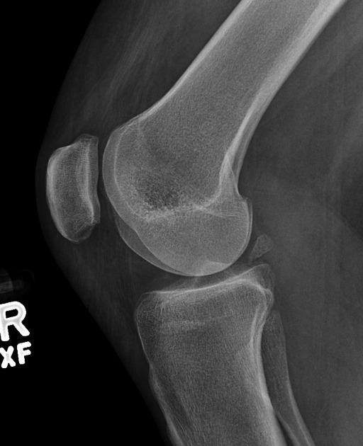
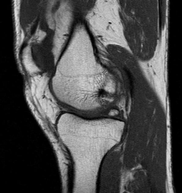
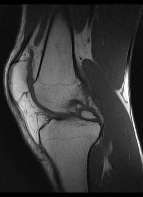
Meniscal ossicles are uncommon, often incidental, findings on radiography and cross-sectional imaging of the knee.
On this page:
Epidemiology
Reported to have a prevalence of 0.15% in the general population 2.
Clinical presentation
- maybe an incidental finding
- may present with intermittent pain
- joint locking is atypical, compared with intra-articular loose bodies
Pathology
The etiology of a meniscal ossicle has not been definitively established, and congenital, traumatic, and degenerative origins have been suggested. Its association with the posterior horn of the medial meniscus may favor a traumatic origin 1.
It consists of cancellous bone with a cartilage interface. There is no fibroblast proliferation or neovascularization 3.
Radiographic findings
Plain radiography / CT
- more often in the posterior horn of the medial meniscus
- should have smooth margins, as opposed to a fracture fragment, and no donor site from the femur or tibia
MRI
The ossicle should follow bone marrow signal on all sequences:
- T1: hyperintense
- T2FS/STIR: hypointense
Treatment and prognosis
If symptomatic, conservative noninterventional therapy is tried first. If this fails, arthroscopic resection can be considered. Interventional therapy may also be considered if the ossicle is associated with other meniscal pathology.


 Unable to process the form. Check for errors and try again.