
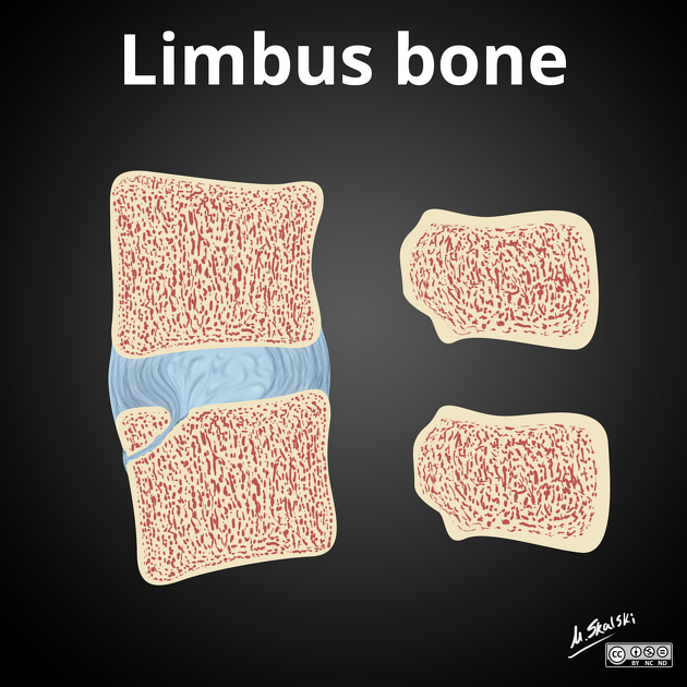
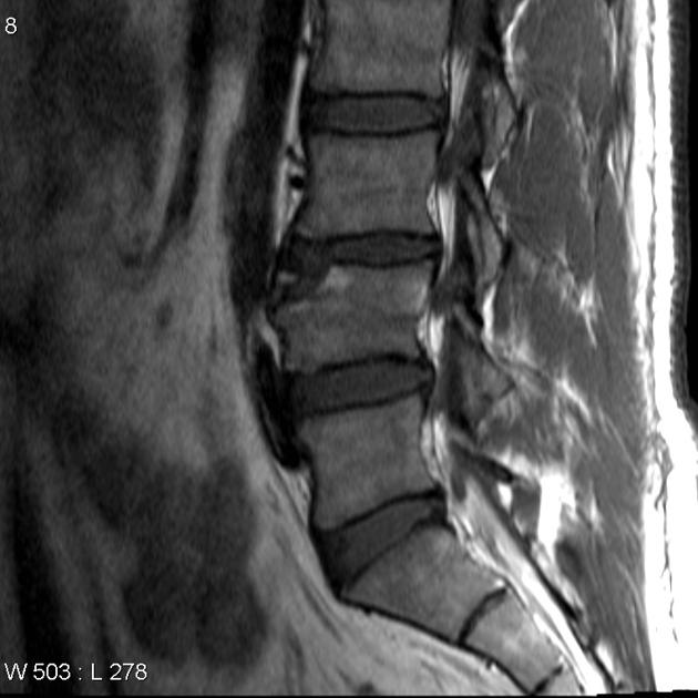
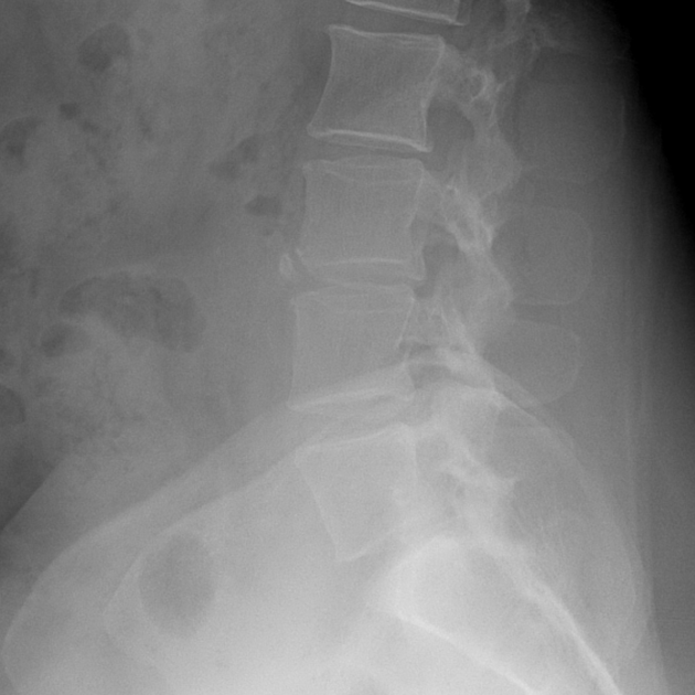
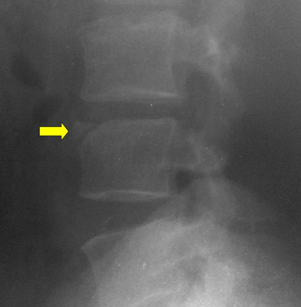
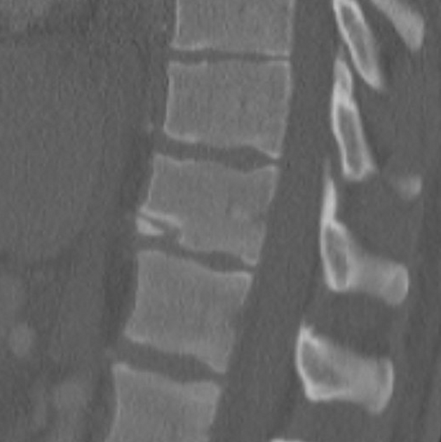
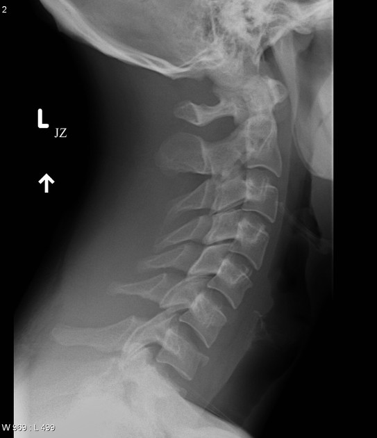
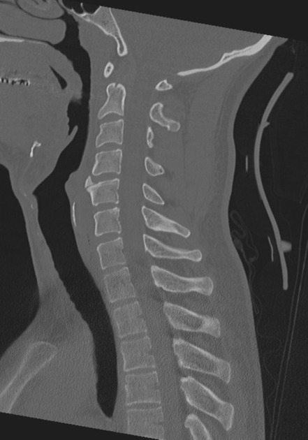
A limbus vertebra is a well-corticated unfused secondary ossification center of the vertebral body, usually of its anterosuperior corner, that occurs secondary to herniation of the nucleus pulposus through the vertebral body endplate beneath the ring apophysis (see ossification of the vertebrae). These are closely related to Schmorl nodes and should not be confused with limbus fractures or infection.
On this page:
Epidemiology
Their formation occurs before the age of 18 years, but often they are seen in older adults.
Clinical presentation
Anterior limbus vertebrae are generally asymptomatic and are detected incidentally. Posterior limbus vertebrae are far less common but have been reported to cause nerve compression.
Radiographic features
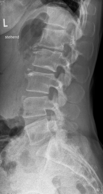
The features of a limbus vertebra are the same on x-rays, CT and MRI.
It should:
be well-corticated (have a sclerotic margin) with a smooth sclerotic adjacent corticated vertebral margin
triangular in shape
occupy the expected location of a normal vertebral body corner
Unlike a fracture, in a limbus vertebra, the 'fragment' of bone will not 'fit' into the adjacent vertebral body defect but rather will usually appear to be too small.
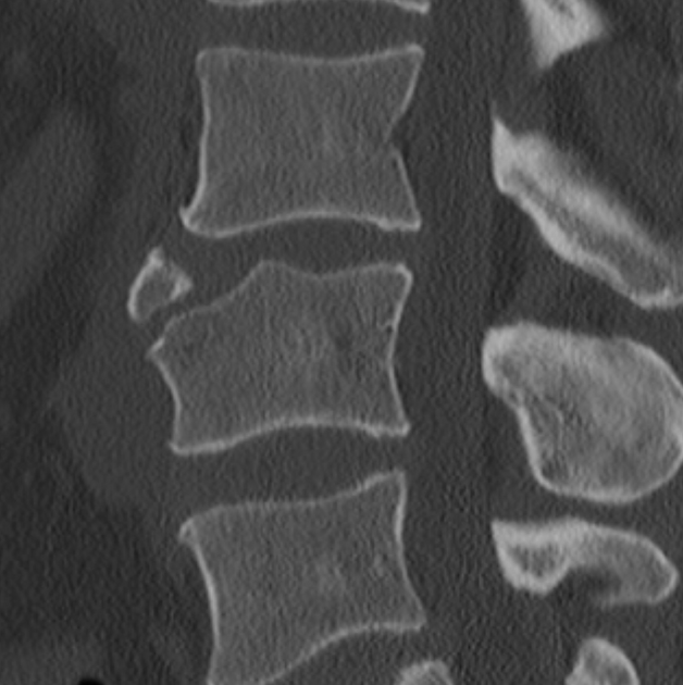
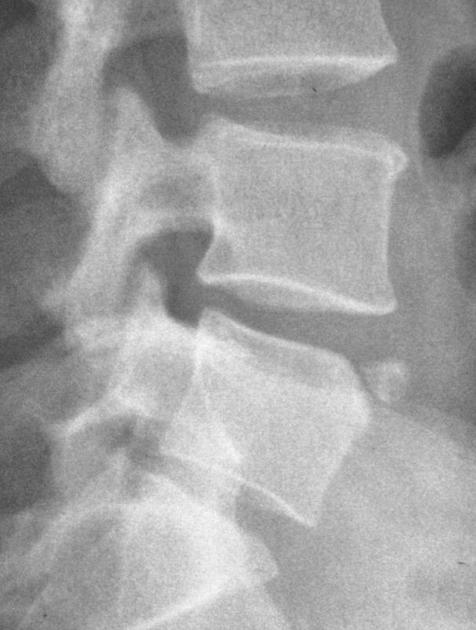
A limbus vertebra is usually encountered in the mid-lumbar spine, although occasionally it may be seen in the thoracic spine. The anterosuperior corner of a single vertebral body in the mid-lumbar spine is the most common presentation. The anteroinferior and posteroinferior corners are seen far less frequently 1.
Discography
Radiography with or without CT or MRI is sufficient for diagnosis. Historically, the diagnosis was confirmed with discography where contrast could be seen extending into the intraosseous herniation of the nucleus pulposus 1.
History and etymology
The term limbus is a direct borrowing from the Latin word meaning fringe, as in the edge of something, or hem 3. Interestingly, limbus vertebra was first described by Schmorl in 1927 4.
Differential diagnosis
Consider
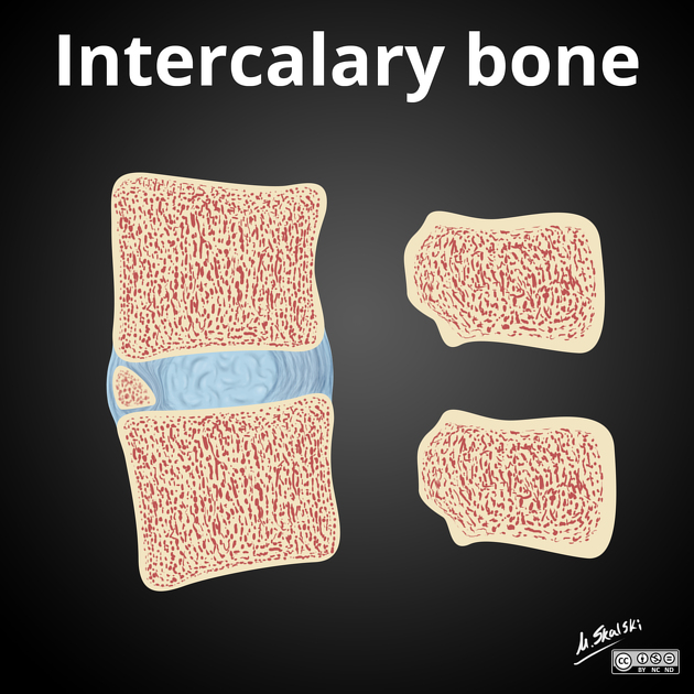
intercalary bone: ossification is in the anterior annular fibers of an intervertebral disc
-
acute fractures: should have an adjacent perivertebral hematoma
teardrop fracture (cervical spine)
degenerative disease of the spine
infection: adjacent cortical loss and soft tissue mass


 Unable to process the form. Check for errors and try again.