
Adrenal insufficiency refers to inadequate secretion of corticosteroids (glucocorticoids and mineralocorticoids).
On this page:
Terminology
It may occur from partial or complete destruction of the adrenal cortex, in which case it is termed primary adrenal insufficiency (also known as Addison disease). Secondary adrenal insufficiency due to lack of stimulation of the gland is a more common etiology overall.
Clinical presentation
Depends on the course of the disease:
acute stage: the patient presents with fever, back pain, hypotension, weakness
chronic stage: progressive lethargy, weakness, cutaneous pigmentation, weight loss
Markers
chemistry: hyponatremia, hyperkalemia, azotemia, hypercalcemia, hypoglycemia
-
adrenocorticotrophic hormone (ACTH) stimulation test
-
primary adrenal insufficiency
cortisol levels fail to rise after synthetic ACTH stimulation due to the destruction of the adrenal cortex
-
secondary adrenal insufficiency
cortisol levels rise after synthetic ACTH stimulation
this indicates decreased ACTH secretion due to pituitary dysfunction
-
Pathology
Primary
idiopathic autoimmune disorders are the most common cause in developed countries (80% of cases) 1
granulomatous disease: tuberculosis (the most common infectious cause in developing countries) and sarcoidosis
neoplasms: metastases (e.g. lung, ovary, kidney, melanoma), lymphoma, and leukemia
adrenal hemorrhage: shock, sepsis (Waterhouse-Friderichsen syndrome), coagulation disorders, antiphospholipid syndrome, trauma 5
systemic fungal infection: histoplasmosis (most common in the southeastern and south-central United States 1)
drugs inhibiting steroidogenesis 7
bilateral adrenalectomy 8
Secondary
suppression of the adrenal axis by endogenous or exogenous glucocorticoids
Time course
The disease course may be either acute, subacute, or chronic 2:
acute: occurs within a few weeks to months and is caused by bilateral adrenal hemorrhage (adrenal apoplexy) or secondary to shock and sepsis or trauma (Addisonian crisis)
subacute disease (adrenalitis): when the disease has been present for less than two years
-
chronic: secondary to chronic autoimmune disorder or chronic granulomatous infection (tuberculosis)
the adrenal glands become atrophic and calcified
Radiographic features
Adrenal insufficiency is a bilateral process that cannot be diagnosed by imaging alone. CT would be the best imaging modality for adrenal assessment but is not necessary for diagnosis.
CT
Imaging features depend on the cause and the course of the disease either acute, subacute, or chronic:
acute: bilateral adrenal hematomas
subacute (adrenalitis): enlargement of both adrenal glands, with necrotic centers and peripheral enhancing rims
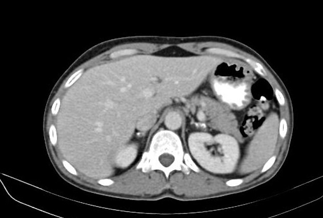
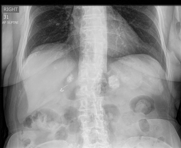
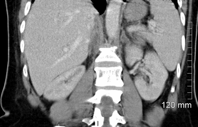
chronic: both adrenal glands appear small and atrophic, associated with calcifications (adrenal calcification) in granulomatous adrenalitis
Treatment and prognosis
acute: glucocorticoid therapy, volume, and electrolyte replacement
chronic: glucocorticoid and mineralocorticoid replacement


 Unable to process the form. Check for errors and try again.