
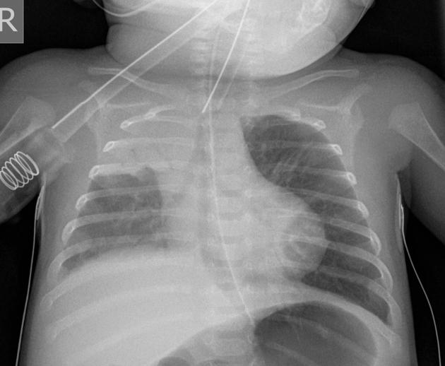
Consolidation
Citation, DOI, disclosures and article data
At the time the article was created Frank Gaillard had no recorded disclosures.
View Frank Gaillard's current disclosuresAt the time the article was last revised Joshua Yap had no financial relationships to ineligible companies to disclose.
View Joshua Yap's current disclosures- Lung consolidation
- Airspace shadowing
- Air-space consolidation
- Air space consolidation
- Airspace opacification
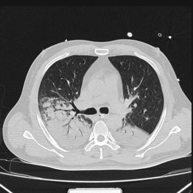
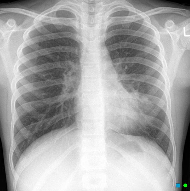
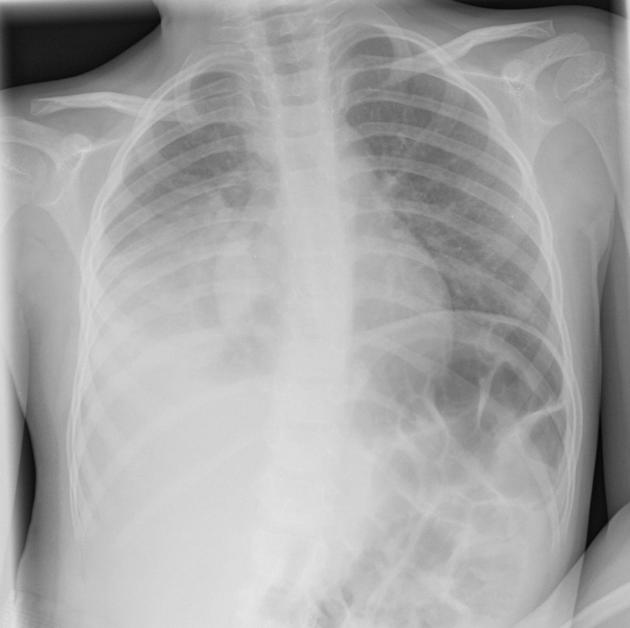
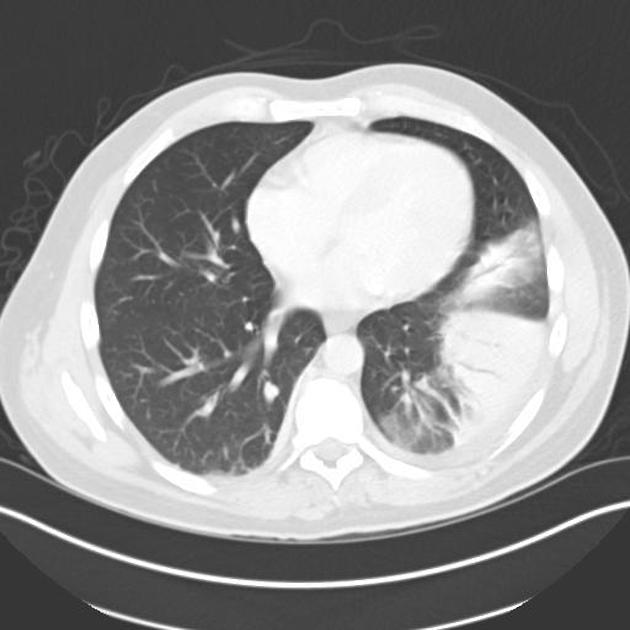
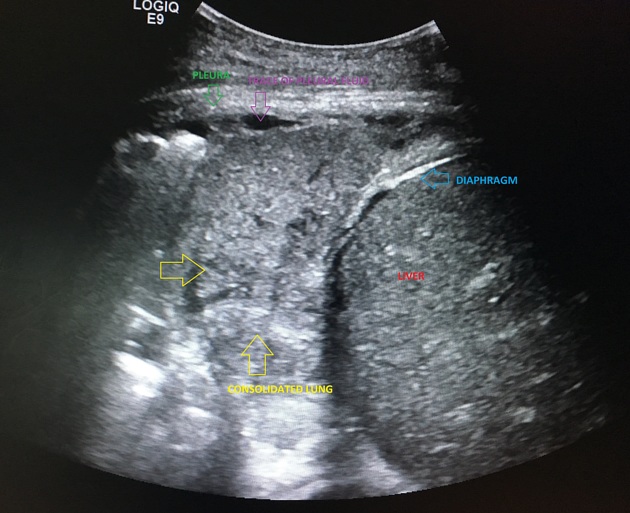
Consolidation describes increased lung attenuation sufficient to obscure bronchial walls and blood vessels (on non-enhanced CT). Patent airways can be identified by the endoluminal gas as an air bronchogram. Consolidation can be caused by any process that evacuates alveolar air such as pneumonia, when air is replaced by inflammatory exudate, or tumour, when air is replaced by tumour cells 3.
On radiographs, consolidation appears as air bronchograms within opacity which is otherwise homogeneous.
Pathology
Aetiology
The opacification is caused by fluid or solid material within the airways that causes a difference in the relative attenuation of the lung:
blood, e.g. pulmonary haemorrhage
cells, e.g. adenocarcinoma
fat, e.g. lipoid pneumonia
gastric contents, e.g. aspiration pneumonia
protein, e.g. alveolar proteinosis
pus, e.g. bacterial pneumonia
transudate, e.g. pulmonary oedema secondary to heart failure
water, e.g. drowning
When considering the likely causes of airspace opacification, it is useful to determine chronicity (by reviewing previous radiographs) and considering laterality.
Additionally, the presence of mediastinal or hilar lymphadenopathy further refines the massive list of differentials:
Patterns of disease
On chest radiography a number of patterns are recognised:
Quiz questions

References
- 1. Kuhlman J, Scatarige J, Fishman E, Zerhouni E, Siegelman S. CT Demonstration of High Attenuation Pleural-Parenchymal Lesions Due to Amiodarone Therapy. J Comput Assist Tomogr. 1987;11(1):160-2. doi:10.1097/00004728-198701000-00034 - Pubmed
- 2. Silva C, Marchiori E, Souza Júnior A, Müller N. Illustrated Brazilian Consensus of Terms and Fundamental Patterns in Chest CT Scans. J Bras Pneumol. 2010;36(1):99-123. doi:10.1590/s1806-37132010000100016 - Pubmed
- 3. Bankier A, MacMahon H, Colby T et al. Fleischner Society: Glossary of Terms for Thoracic Imaging. Radiology. 2024;310(2):e232558. doi:10.1148/radiol.232558 - Pubmed
Incoming Links
- Pulmonary nocardiosis
- Hemithorax white-out (differential)
- Lung consolidation (mnemonic)
- Pulmonary Mycobacterium chelonae infection
- COVID-19
- Acute respiratory distress syndrome
- Chronic unilateral airspace opacification (differential)
- Coccidioidomycosis
- Pulmonary infection
- Pulmonary mucormycosis
- Pulmonary coccidioidomycosis
- Bronchopneumonia
- Fluid bronchogram sign
- Hypersensitivity pneumonitis
- Cytomegalovirus pulmonary infection
- Necrotising pneumonia
- Rituximab-induced interstitial lung disease
- Pulmonary pseudomonas aeruginosa infection
- Hospital-acquired pneumonia
- Honeycombing (lungs)
- Lung adenocarcinoma presenting as pure ground-glass nodule
- Pectus excavatum with COVID19
- Right middle and lower lobe pneumonia - Mycoplasma
- Pulmonary lacerations
- Spine sign
- White-out hemithorax
- Pneumonia
- Peripheral pulmonary consolidation
- Pulmonary infarction
- Pneumonia
- Hydatid cyst - calcified
- Pulmonary tuberculosis with tuberculous orchitis
- Pulmonary edema
- Invasive mucinous adenocarcinoma of the lung mimicking pneumonia
- Atoll sign in COVID-19 pneumonia
- Lung adenocarcinoma presenting as consolidation
- Cardiogenic pulmonary edema
- Cardiogenic pulmonary oedema - unilateral
- COVID-19 pneumonia
- H1N1 pneumonia
Related articles: Airspace opacification
- airspace opacification
- differential diagnoses of airspace opacification
- lobar consolidation
-
atelectasis
- mechanism-based
- morphology-based
- lobar lung collapse
Related articles: Chest
- imaging techniques
-
chest radiograph
- radiography
-
approach
- ABCDE
- ABCDEFGHI
- congenital heart disease
- medical devices in the thorax
- common lines and tubes
- nasogastric tubes
- endotracheal tubes
- central venous catheters
- oesophageal temperature probe
- tracheostomy tube
- pleural catheters
- cardiac conduction devices
- prosthetic heart valve
- review areas
-
airspace opacification
- differential diagnoses of airspace opacification
- lobar consolidation
-
atelectasis
- mechanism-based
- morphology-based
- lobar lung collapse
- chest x-ray in the exam setting
- cardiomediastinal contour
- chest radiograph zones
- tracheal air column
- fissures
- normal chest x-ray appearance of the diaphragm
- nipple shadow
-
lines and stripes
- anterior junction line
- posterior junction line
- right paratracheal stripe
- left paratracheal stripe
- posterior tracheal stripe/tracheo-oesophageal stripe
- posterior wall of bronchus intermedius
- right paraspinal line
- left paraspinal line
- aortic-pulmonary stripe
- aortopulmonary window
- azygo-oesophageal recess
- spaces
- signs
- air bronchogram
- big rib sign
- Chang sign
- Chen sign
- coin lesion
- continuous diaphragm sign
- dense hilum sign
- double contour sign
- egg-on-a-string sign
- extrapleural sign
- finger in glove sign
- flat waist sign
- Fleischner sign
- ginkgo leaf sign
- Golden S sign
- Hampton hump
- haystack sign
- hilum convergence sign
- hilum overlay sign
- Hoffman-Rigler sign
- holly leaf sign
- incomplete border sign
- juxtaphrenic peak sign
- Kirklin sign
- medial stripe sign
- melting ice cube sign
- more black sign
- Naclerio V sign
- Palla sign
- pericardial fat tag sign
- Shmoo sign
- silhouette sign
- snowman sign
- spinnaker sign
- steeple sign
- straight left heart border sign
- third mogul sign
- tram-track sign
- walking man sign
- water bottle sign
- wave sign
- Westermark sign
- HRCT
-
chest radiograph
- airways
- bronchitis
- small airways disease
-
bronchiectasis
- broncho-arterial ratio
- related conditions
- differentials by distribution
- narrowing
-
tracheal stenosis
- diffuse tracheal narrowing (differential)
-
bronchial stenosis
- diffuse airway narrowing (differential)
-
tracheal stenosis
- diverticula
- pulmonary oedema
-
interstitial lung disease (ILD)
- Anti-Jo-1 antibody-positive interstitial lung disease
- drug-induced interstitial lung disease
-
hypersensitivity pneumonitis
- acute hypersensitivity pneumonitis
- subacute hypersensitivity pneumonitis
- chronic hypersensitivity pneumonitis
- aetiology
- bird fancier's lung: pigeon fancier's lung
- farmer's lung
- cheese workers' lung
- bagassosis
- mushroom worker’s lung
- malt worker’s lung
- maple bark disease
- hot tub lung
- wine maker’s lung
- woodsman’s disease
- thatched roof lung
- tobacco grower’s lung
- potato riddler’s lung
- summer-type pneumonitis
- dry rot lung
- machine operator’s lung
- humidifier lung
- shower curtain disease
- furrier’s lung
- miller’s lung
- lycoperdonosis
- saxophone lung
-
idiopathic interstitial pneumonia (mnemonic)
- acute interstitial pneumonia (AIP)
- cryptogenic organising pneumonia (COP)
- desquamative interstitial pneumonia (DIP)
- non-specific interstitial pneumonia (NSIP)
- idiopathic pleuroparenchymal fibroelastosis
- lymphoid interstitial pneumonia (LIP)
- respiratory bronchiolitis–associated interstitial lung disease (RB-ILD)
- usual interstitial pneumonia / idiopathic pulmonary fibrosis (UIP/IPF)
-
pneumoconioses
- fibrotic
- non-fibrotic
-
lung cancer
-
non-small-cell lung cancer
-
adenocarcinoma
- pre-invasive tumours
- minimally invasive tumours
- invasive tumours
- variants of invasive carcinoma
- described imaging features
- adenosquamous carcinoma
- large cell carcinoma
- primary sarcomatoid carcinoma of the lung
- squamous cell carcinoma
- salivary gland-type tumours
-
adenocarcinoma
- pulmonary neuroendocrine tumours
- preinvasive lesions
-
lung cancer invasion patterns
- tumour spread through air spaces (STAS)
- presence of non-lepidic patterns such as acinar, papillary, solid, or micropapillary
- myofibroblastic stroma associated with invasive tumour cells
- pleural invasion
- vascular invasion
- tumours by location
- benign neoplasms
- pulmonary metastases
- lung cancer screening
- lung cancer staging
-
non-small-cell lung cancer


 Unable to process the form. Check for errors and try again.