
Anterior pituitary
Citation, DOI, disclosures and article data
At the time the article was created Craig Hacking had no recorded disclosures.
View Craig Hacking's current disclosuresAt the time the article was last revised Francis Deng had no recorded disclosures.
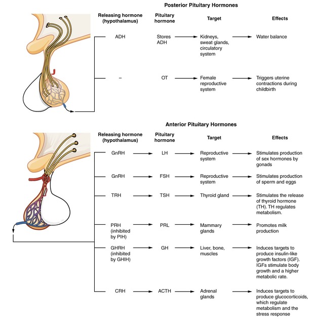
View Francis Deng's current disclosuresThe anterior pituitary (or adenohypophysis) is by far the largest part of the pituitary gland, and is responsible for synthesis and release of most pituitary hormones (with the exception of oxytocin and antidiuretic hormone (ADH) which are released by the posterior pituitary).
It consists of 3 parts:
Pars distalis
The pars distalis is the largest part of the pituitary gland. It forms from the anterior wall of Rathke pouch. It is composed of cords of epithelial cells individually specialised to secrete tropic hormones acting on target organs:
- growth hormone (somatotropin)
- thyroid stimulating hormone (TSH)
- adrenocorticotropic hormone (ACTH)
- follicular stimulating hormone (FSH)
- leutinizing hormone (LH)
- prolactin
Pars tuberalis
The pars tuberalis is the part of the adenohypophysis which surrounds the anterior aspect of the infundibular stalk.
Pars intermedia
The pars intermedia in a thin layer of epithelial cells located between pars distalis and neurohypophysis. It arises from the posterior wall of Rathke pouch and contains vestigial lumina of Rathke pouch which appear as narrow vesicles of variable length. These may give rise to Rathke cleft cysts (also known as pars intermedia cysts 2-3).
For relations and blood supply, please see the main pituitary gland article here.
References
- 1. Moore KL, Agur AMR, Dalley AF. Clinically oriented anatomy. LWW. ISBN:1451119453. Read it at Google Books - Find it at Amazon
- 2. Byun WM, Kim OL, Kim D. MR imaging findings of Rathke's cleft cysts: significance of intracystic nodules. AJNR Am J Neuroradiol. 2000;21 (3): 485-8. AJNR Am J Neuroradiol (full text) - Pubmed citation
- 3. Crenshaw WB, Chew FS. Rathke's cleft cyst. AJR Am J Roentgenol. 1992;158 (6): 1312. AJR Am J Roentgenol (citation) - Pubmed citation
- 4. Last's anatomy, regional and applied. Churchill Livingstone. ISBN:044304662X. Read it at Google Books - Find it at Amazon
- 5. Butler P, Mitchell A, Healy JC. Applied Radiological Anatomy. Cambridge University Press. (2012) ISBN:0521766664. Read it at Google Books - Find it at Amazon
Incoming Links
Related articles: Anatomy: Brain
-
brain
- grey matter
- white matter
-
cerebrum
-
cerebral hemisphere (telencephalon)
- cerebral lobes and gyri
- frontal lobe
- parietal lobe
-
occipital lobe
- occipital pole
- lingual gyrus
- fusiform gyrus (Brodmann area 37)
- calcarine (visual) cortex
- cuneus
- temporal lobe
- basal forebrain
- limbic system
- insula
-
cerebral sulci and fissures (A-Z)
- calcarine fissure
- callosal sulcus
- central (Rolandic) sulcus
- cingulate sulcus
- collateral sulcus
- inferior frontal sulcus
- inferior occipital sulcus
- inferior temporal sulcus
- interhemispheric fissure
- intraparietal sulcus
- lateral (Sylvian) sulcus
- lateral occipital sulcus
- marginal sulcus
- occipitotemporal sulcus
- olfactory sulcus
- paracentral sulcus
- paraolfactory sulcus
- parieto-occipital fissure
- posterior parolfactory sulcus
- precentral sulcus
- preoccipital notch
- postcentral sulcus
- rhinal sulcus
- rostral sulcus
- subparietal sulcus
- superior frontal sulcus
- superior occipital sulcus
- superior temporal sulcus
- cortical histology
- cerebral lobes and gyri
- white matter tracts
- deep grey matter
-
pituitary gland
- posterior pituitary and stalk (part of diencephalon)
- anterior pituitary
- inferior hypophyseal arterial circle
- diencephalon
-
cerebral hemisphere (telencephalon)
-
brainstem
- midbrain (mesencephalon)
- pons (part of metencephalon)
- medulla oblongata (myelencephalon)
- white matter
-
grey matter
- non-cranial nerve
-
cranial nerve nuclei
- oculomotor nucleus
- Edinger-Westphal nucleus
- trochlear nucleus
- motor nucleus of CN V
- mesencephalic nucleus of CN V
- main sensory nucleus of CN V
- spinal nucleus of CN V
- abducent nucleus
- facial nucleus
- superior salivatory nucleus
- cochlear nuclei
- vestibular nuclei
- inferior salivatory nucleus
- solitary tract nucleus
- ambiguus nucleus
- dorsal vagal motor nucleus
- hypoglossal nucleus
-
cerebellum (part of metencephalon)
- vermis
- cerebellar hemisphere
- cerebellar peduncles
- cranial meninges (meninx primitiva)
- CSF spaces
-
cranial nerves (mnemonic)
- olfactory nerve (CN I)
- optic nerve (CN II)
- oculomotor nerve (CN III)
- trochlear nerve (CN IV)
- trigeminal nerve (CN V) (mnemonic)
- abducens nerve (CN VI)
- facial nerve (CN VII) (segments mnemonic | branches mnemonic)
-
vestibulocochlear nerve (CN VIII)
- vestibular ganglion (Scarpa's ganglion)
- glossopharyngeal nerve (CN IX)
- vagus nerve (CN X)
- spinal accessory nerve (CN XI)
- hypoglossal nerve (CN XII)
- functional neuroanatomy
- CNS development
- cerebral vascular supply
- arteries
- vascular territories
-
circle of Willis
- internal carotid artery (ICA) (segments)
- vertebral artery
-
normal variants
- intracranial arterial fenestration
- internal carotid artery (ICA)
- anterior cerebral artery (ACA)
- middle cerebral artery (MCA)
- posterior cerebral artery (PCA)
- basilar artery
- persistent carotid-vertebrobasilar artery anastomoses (mnemonic)
- vertebral artery
- ophthalmic artery
-
cerebral venous system
-
dural venous sinuses
- basilar venous plexus
- cavernous sinus (mnemonic)
- clival diploic veins
- inferior petro-occipital vein
- inferior petrosal sinus
- inferior sagittal sinus
- intercavernous sinus
- internal carotid artery venous plexus of Rektorzik
- jugular bulb
- marginal sinus
- occipital sinus
- sigmoid sinus
- sphenoparietal sinus
- straight sinus
- superior petrosal sinus
- superior sagittal sinus
- torcula herophili
- transverse sinus
-
cerebral veins
-
superficial veins of the brain
- superior cerebral veins (superficial cerebral veins)
- inferior cerebral veins
- superficial middle cerebral vein
- superior anastomotic vein (of Trolard)
- inferior anastomotic vein (of Labbe)
-
superficial veins of the brain
-
deep veins of the brain
- great cerebral vein (of Galen)
- venous circle of Trolard
- normal variants
-
dural venous sinuses
- arteries
- glymphatic pathway


 Unable to process the form. Check for errors and try again.