
This is a basic article for medical students and other non-radiologists
Appendicitis occurs when there is inflammation of the vermiform appendix. It is a very common condition and is a major cause of abdominal surgery in young patients. CT is the most sensitive modality to detect appendicitis although its use should be limited because of the radiation dose required and ultrasound should be employed as first-line where possible.
On this page:
Reference article
This is a summary article; read more in our article on appendicitis.
Summary
-
anatomy
-
epidemiology
typically children and young adults
peak incidence 2nd to 3rd decade of life
-
presentation
initial periumbilical pain with fever, nausea and vomiting
progresses to localised right iliac fossa pain
-
pathophysiology
-
obstruction of the appendiceal lumen
fluid accumulation, infection, venous congestion, ischaemia/necrosis
-
causes
lymphoid hyperplasia (60%)
appendicolith (33%)
rare: foreign body, Crohn's disease, tumour
-
-
investigation
US is often all that is required (quick, dynamic and no ionising radiation)
cross-sectional imaging is more sensitive (CT and MRI)
-
treatment
appendicectomy (laparoscopic or open)
-
prognosis
prognosis is very good and mortality very low (0.1%)
-
complications include:
perforation and abscess formation
generalised peritonitis
Role of imaging
confirm appendicitis
assess for peri-appendiceal collection
assess for perforation
determine if there is another cause for symptoms
Radiographic features
Ultrasound
Graded compression, and uses the linear probe over the site of maximal tenderness, with gradually increasing pressure to displace normal overlying bowel gas. Always used in young patients because of the lack of ionising radiation.
distended appendix
surrounding (echogenic) inflamed fat
thickening (oedema) and then later, thinning (pre-rupture) of the appendix wall
increased appendix wall vascularity
collections (hypoechoic areas) around the appendix
In one meta-analysis, ultrasound has sensitivity and specificity of 69% and 81%, respectively, for the diagnosis of acute appendicitis.1
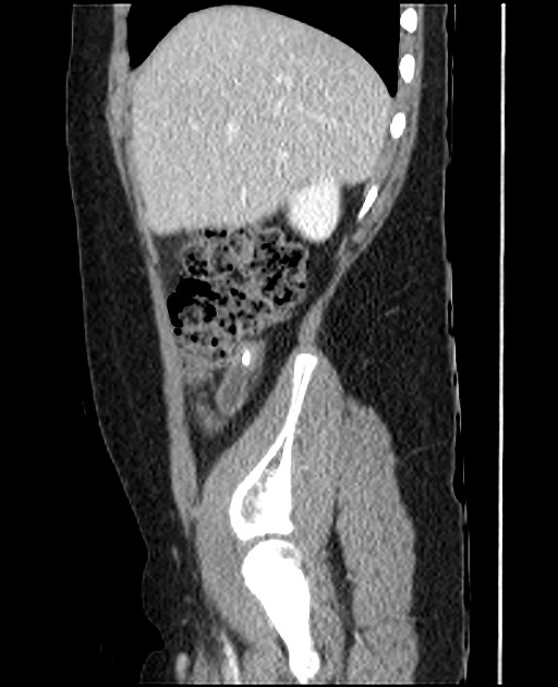
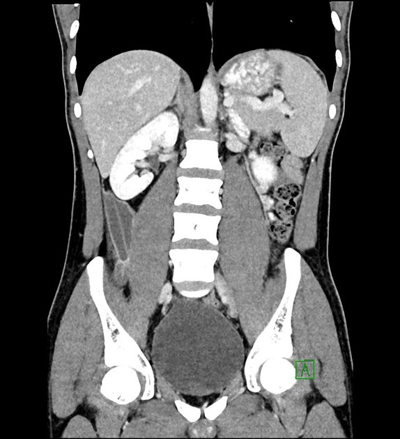
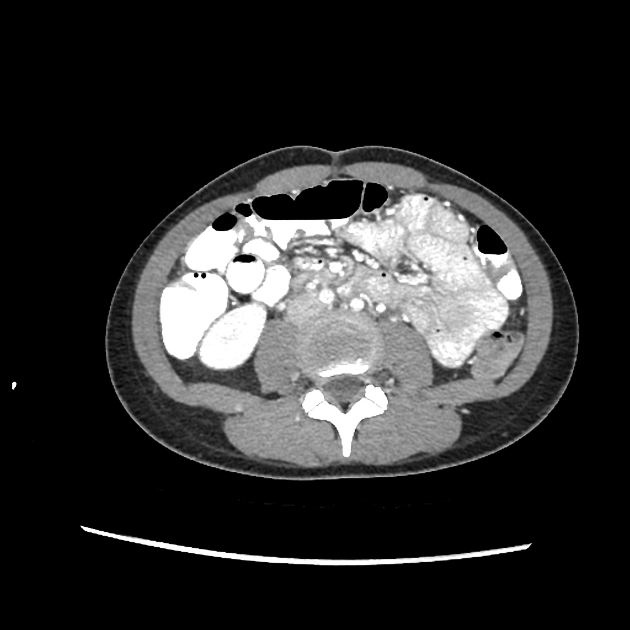
CT
CT is highly sensitive (94-98%) and specific (up to 97%) for the diagnosis of acute appendicitis and allows other causes of abdominal pain to be diagnosed. Usually performed with IV contrast (no oral contrast required).
dilated appendix with a distended lumen ( >6 mm diameter)
thickened and enhancing wall
thickening of the caecal apex (up to 80%)
peri-appendiceal inflammation (stranding of the adjacent fat)
extraluminal fluid
inflammatory phlegmon
abscess formation
appendicolith may also be identified
MRI
In one study, the sensitivity and specificity of MRI for the diagnosis of acute appendicitis are 96% and 96%, respectively. The appearance of appendicitis on MRI has many similarities to the CT appearance 2.


 Unable to process the form. Check for errors and try again.