
Arachnoid granulation
Citation, DOI, disclosures and article data
At the time the article was created Frank Gaillard had no recorded disclosures.
View Frank Gaillard's current disclosuresAt the time the article was last revised Liz Silverstone had no financial relationships to ineligible companies to disclose.
View Liz Silverstone's current disclosures- Pacchionian bodies
- Pacchionian granulations
- Pacchionian granulation
- Arachnoid granulations
- Arachnoid villi
- Pacchion's granulations
- Pacchion granulations
- Pacchion granulation
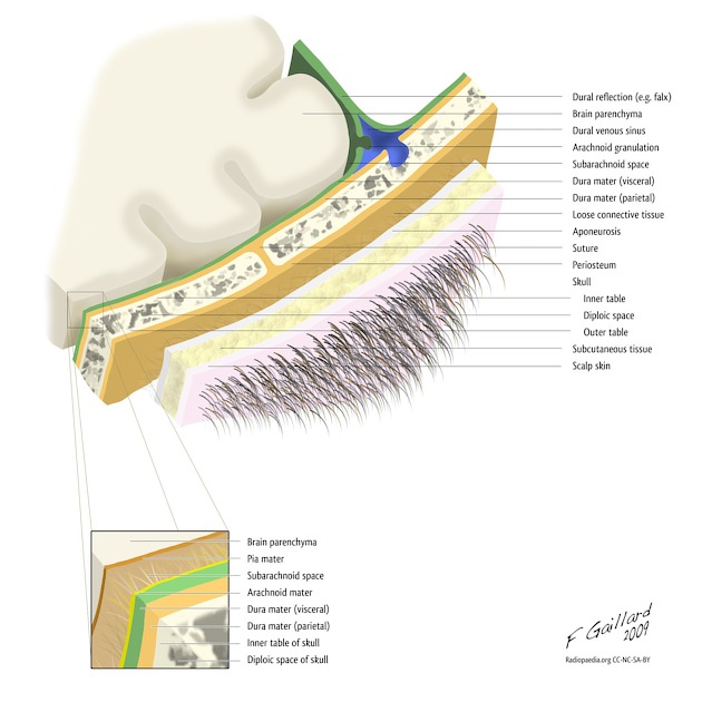
Arachnoid granulations, also known as Pacchionian granulations, are projections of the arachnoid membrane (villi) into the dural sinuses that allow CSF to pass from the subarachnoid space into the venous system.
On this page:
Epidemiology
They increase in size and number with age and are seen in approximately two-thirds of patients.
Pathology
Location
They most frequently occur in a parasagittal location with the transverse and superior sagittal sinuses being the most common locations. The granulations typically occur next to the entrance of a superficial draining cortical vein into a sinus (similar to colonic diverticula occurring next to penetrating vessels).
Radiographic features
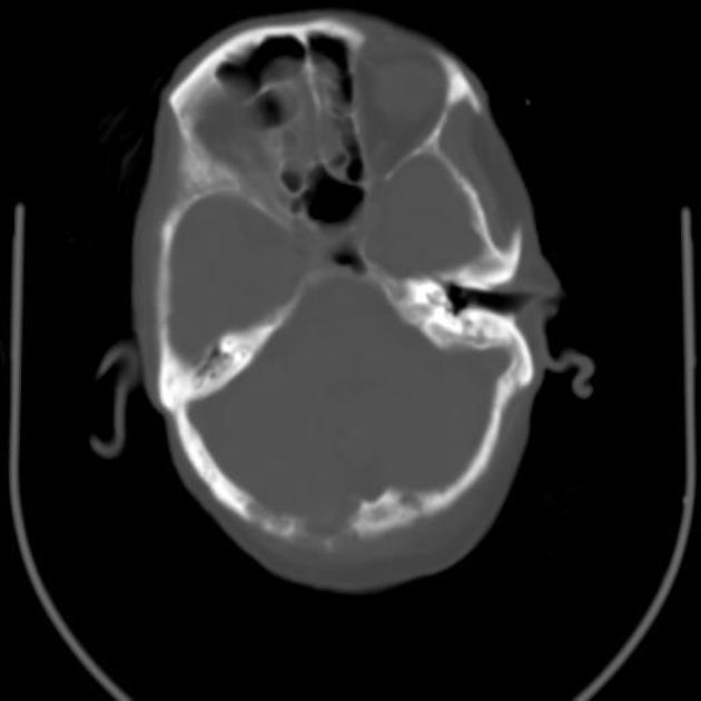
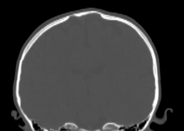
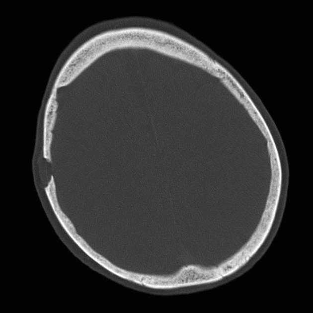
They are most commonly encountered in radiological practice as incidental osteolytic, sharply circumscribed indolent-appearing lucencies on skull CT or x-rays, or a filling defect in dural venous sinuses, which can be mistaken for dural venous thrombosis.
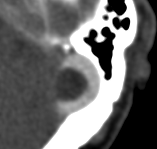
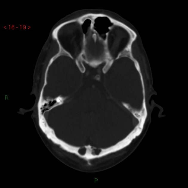
CT
The granulations are typically of CSF density and protrude into the calvaria or a dural venous sinus causing a filling defect. They may simulate a dural venous sinus thrombosis but are usually easy differentiated given their round well-defined shape and classic location.
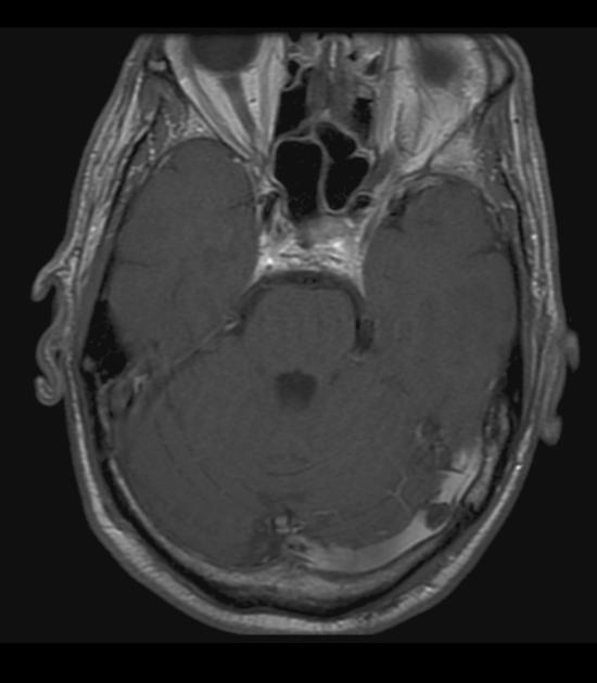
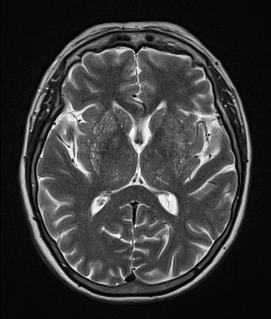
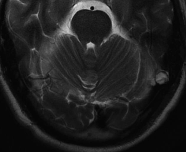
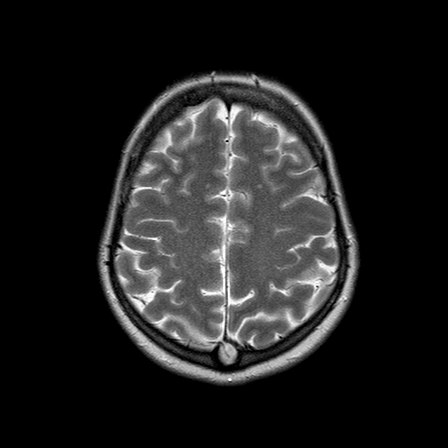
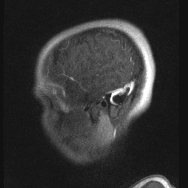
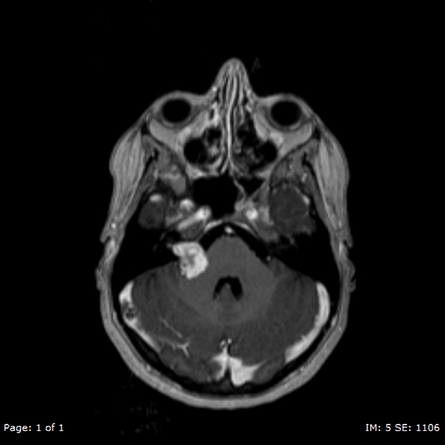
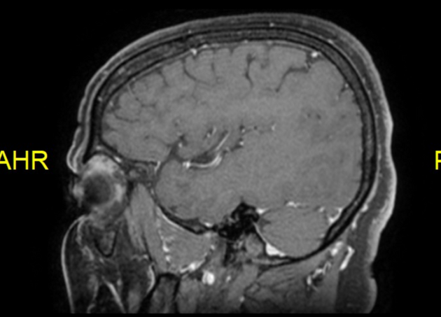
MRI
Signal characteristics are generally those of CSF 2:
T1: low signal intensity
T2: high signal intensity; iso- or even slightly hyperintense to CSF
FLAIR: should attenuate
T1 C+ (Gd): no enhancement
Giant granulations (>10 mm) may show atypical MRI signal characteristics, with higher T1 and T2 signal than CSF and incomplete FLAIR signal suppression 5. Rarely these may cause sinus obstruction 6.
ADVERTISEMENT: Supporters see fewer/no ads
History and etymology
Arachnoid granulations are named after Antonio Pacchioni (1665-1726), an Italian physician, who wrote extensively on the anatomy of the dura mater, and provided the first written description of his eponymous granulations in 1705 in the Dissertatio Epistolaris de Glandulis Conglobatis Durae Meningis Humanae, one of his monographs 1.
Differential diagnosis
Considerations include
On CT if there is lytic erosion of the bone also consider
small calvarial metastases/myeloma/plasmacytoma etc.
or rarely
intradiploic epidermoid - often centred towards the mid to outer margins of the skull
See also
References
- 1. Brunori A, Vagnozzi R, Giuffrè R. Antonio Pacchioni (1665-1726): early studies of the dura mater. (1993) Journal of neurosurgery. 78 (3): 515-8. doi:10.3171/jns.1993.78.3.0515 - Pubmed
- 2. Ikushima I, Korogi Y, Makita O et-al. MRI of arachnoid granulations within the dural sinuses using a FLAIR pulse sequence. Br J Radiol. 1999;72 (863): 1046-51. Br J Radiol (abstract) - Pubmed citation
- 3. Lu CX, Du Y, Xu XX et-al. Multiple occipital defects caused by arachnoid granulations: Emphasis on T2 mapping. World J Radiol. 2012;4 (7): 341-4. doi:10.4329/wjr.v4.i7.341 - Free text at pubmed - Pubmed citation
- 4. Rodallec MH, Krainik A, Feydy A et-al. Cerebral venous thrombosis and multidetector CT angiography: tips and tricks. Radiographics. 2006;26 Suppl 1 : S5-18. doi:10.1148/rg.26si065505 - Pubmed citation
- 5. Trimble CR, Harnsberger HR, Castillo M et-al. "Giant" arachnoid granulations just like CSF?: NOT!!. AJNR Am J Neuroradiol. 2010;31 (9): 1724-8. doi:10.3174/ajnr.A2157 - Pubmed citation
- 6. Kan P, Stevens EA, Couldwell WT. Incidental giant arachnoid granulation. AJNR Am J Neuroradiol. 2006;27 (7): 1491-2. Pubmed citation
Incoming Links
- Benign enlargement of the subarachnoid space in infancy
- Transverse sinus stenosis
- Venous lacunae (skull)
- CSF flow studies
- Choroid plexus
- Dural venous sinus cyst
- Communicating hydrocephalus
- Normal intracranial calcifications
- Cerebral venous thrombosis
- Encephalocele
- Dural venous sinus thrombosis
- CT cerebral venography (protocol)
- Congenital calvarial defects
- Temporal encephalocele
- Arachnoid mater
- Aberrant arachnoid granulations
- Solitary lucent skull lesion
- Cerebrospinal fluid
- Lytic skull lesion
- Brain herniation into arachnoid granulation
- Brain herniation into arachnoid granulation
- Arachnoid granulation
- Brain herniation into giant arachnoid granulation of the sigmoid sinus
- Arachnoid granulation
- Arachnoid granulation
- Dural venous sinus thrombosis
- Central neurocytoma
- Posterior fossa arachnoid cyst - large
- Arachnoid granulations - typical
- Giant arachnoid granulation
- Arachnoid granulation
- Dural venous sinus thrombosis with hemorrhagic infarction
- Arachnoid granulation
- Arachnoid granulation
- Giant arachnoid granulation
- Dural venous sinus cyst
- Giant arachnoid granulation
- Arachnoid granulation
- Arachnoid granulation
- Intraosseous arachnoid granulations
Related articles: Anatomy: Brain
-
brain
- grey matter
- white matter
-
cerebrum
-
cerebral hemisphere (telencephalon)
- cerebral lobes and gyri
- frontal lobe
- parietal lobe
-
occipital lobe
- occipital pole
- lingual gyrus
- fusiform gyrus (Brodmann area 37)
- calcarine (visual) cortex
- cuneus
- temporal lobe
- basal forebrain
- limbic system
- insula
-
cerebral sulci and fissures (A-Z)
- calcarine fissure
- callosal sulcus
- central (Rolandic) sulcus
- cingulate sulcus
- collateral sulcus
- inferior frontal sulcus
- inferior occipital sulcus
- inferior temporal sulcus
- interhemispheric fissure
- intraparietal sulcus
- lateral (Sylvian) sulcus
- lateral occipital sulcus
- marginal sulcus
- occipitotemporal sulcus
- olfactory sulcus
- paracentral sulcus
- paraolfactory sulcus
- parieto-occipital fissure
- posterior parolfactory sulcus
- precentral sulcus
- preoccipital notch
- postcentral sulcus
- rhinal sulcus
- rostral sulcus
- subparietal sulcus
- superior frontal sulcus
- superior occipital sulcus
- superior temporal sulcus
- cortical histology
- cerebral lobes and gyri
- white matter tracts
- deep grey matter
-
pituitary gland
- posterior pituitary and stalk (part of diencephalon)
- anterior pituitary
- inferior hypophyseal arterial circle
- diencephalon
-
cerebral hemisphere (telencephalon)
-
brainstem
- midbrain (mesencephalon)
- pons (part of metencephalon)
- medulla oblongata (myelencephalon)
- white matter
-
grey matter
- non-cranial nerve
-
cranial nerve nuclei
- oculomotor nucleus
- Edinger-Westphal nucleus
- trochlear nucleus
- motor nucleus of CN V
- mesencephalic nucleus of CN V
- main sensory nucleus of CN V
- spinal nucleus of CN V
- abducent nucleus
- facial nucleus
- superior salivatory nucleus
- cochlear nuclei
- vestibular nuclei
- inferior salivatory nucleus
- solitary tract nucleus
- ambiguus nucleus
- dorsal vagal motor nucleus
- hypoglossal nucleus
-
cerebellum (part of metencephalon)
- vermis
- cerebellar hemisphere
- cerebellar peduncles
- cranial meninges (meninx primitiva)
- CSF spaces
-
cranial nerves (mnemonic)
- olfactory nerve (CN I)
- optic nerve (CN II)
- oculomotor nerve (CN III)
- trochlear nerve (CN IV)
- trigeminal nerve (CN V) (mnemonic)
- abducens nerve (CN VI)
- facial nerve (CN VII) (segments mnemonic | branches mnemonic)
-
vestibulocochlear nerve (CN VIII)
- vestibular ganglion (Scarpa's ganglion)
- glossopharyngeal nerve (CN IX)
- vagus nerve (CN X)
- spinal accessory nerve (CN XI)
- hypoglossal nerve (CN XII)
- functional neuroanatomy
- CNS development
- cerebral vascular supply
- arteries
- vascular territories
-
circle of Willis
- internal carotid artery (ICA) (segments)
- vertebral artery
-
normal variants
- intracranial arterial fenestration
- internal carotid artery (ICA)
- anterior cerebral artery (ACA)
- middle cerebral artery (MCA)
- posterior cerebral artery (PCA)
- basilar artery
- persistent carotid-vertebrobasilar artery anastomoses (mnemonic)
- vertebral artery
- ophthalmic artery
-
cerebral venous system
-
dural venous sinuses
- basilar venous plexus
- cavernous sinus (mnemonic)
- clival diploic veins
- inferior petro-occipital vein
- inferior petrosal sinus
- inferior sagittal sinus
- intercavernous sinus
- internal carotid artery venous plexus of Rektorzik
- jugular bulb
- marginal sinus
- occipital sinus
- sigmoid sinus
- sphenoparietal sinus
- straight sinus
- superior petrosal sinus
- superior sagittal sinus
- torcula herophili
- transverse sinus
-
cerebral veins
-
superficial veins of the brain
- superior cerebral veins (superficial cerebral veins)
- inferior cerebral veins
- superficial middle cerebral vein
- superior anastomotic vein (of Trolard)
- inferior anastomotic vein (of Labbe)
-
superficial veins of the brain
-
deep veins of the brain
- great cerebral vein (of Galen)
- venous circle of Trolard
- normal variants
-
dural venous sinuses
- arteries
- glymphatic pathway


 Unable to process the form. Check for errors and try again.