Citation, DOI, disclosures and article data
Citation:
Gaillard F, Sharma R, Mahsoub M, et al. Bilateral middle cerebellar peduncle lesions. Reference article, Radiopaedia.org (Accessed on 17 Mar 2025) https://doi.org/10.53347/rID-13017
Bilateral lesions of the middle cerebellar peduncles, resulting in the middle cerebellar peduncle sign, are uncommon and can be seen either in isolation (rare) or along with other regions of involvement.
Despite their relative rarity, they have a fairly long list of potential causes (see below) 1-4. Among these, neurodegenerative diseases are probably most common.
Pathology
The middle cerebellar peduncles are composed of white matter that connects the cerebellar hemispheres to the contralateral pontine nuclei 5. Therefore, many of the causes of lesions in this location are related to white matter pathology. They are supplied by branches of the anterior inferior cerebellar artery and the superior cerebellar artery.
For additional discussion of region anatomy refer to middle cerebellar peduncles.
Etiology
Most of the entities listed do not usually cause isolated bilateral middle cerebellar peduncular lesions and as such care must be taken in identifying other areas of involvement.
Radiographic features
Although potentially visible on CT, in most instances this is an MRI feature.
MRI
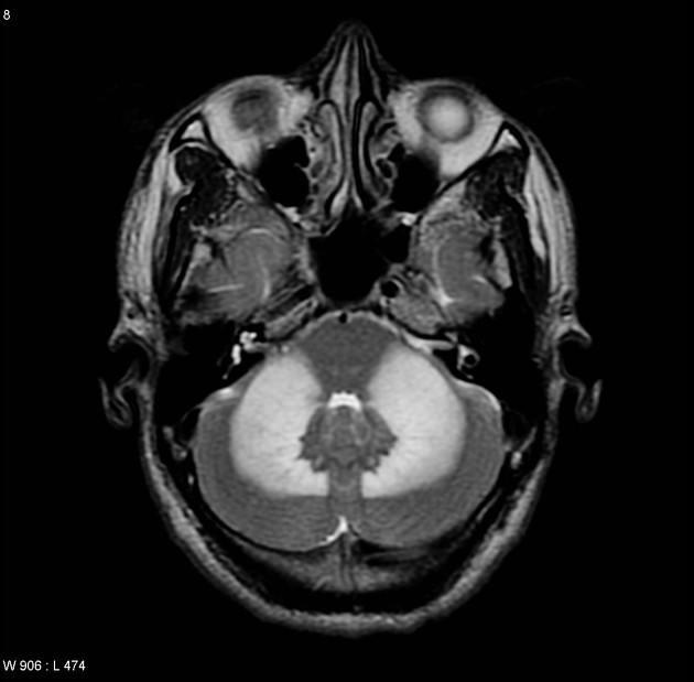
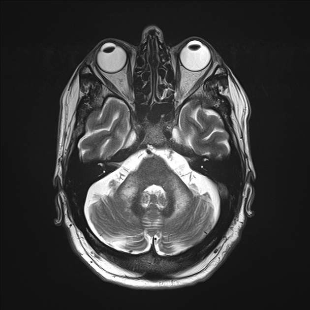
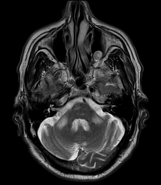
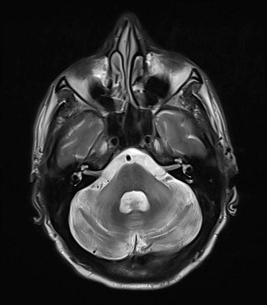
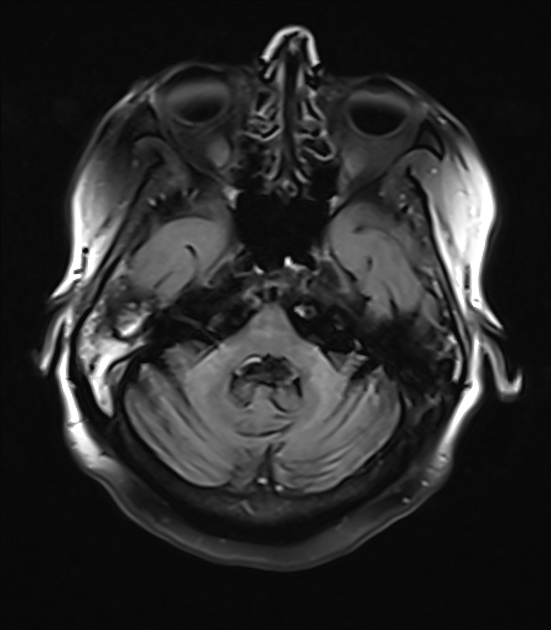
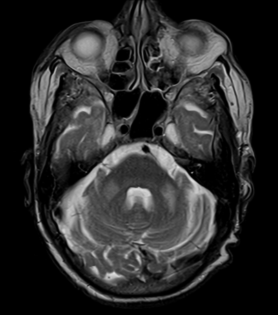
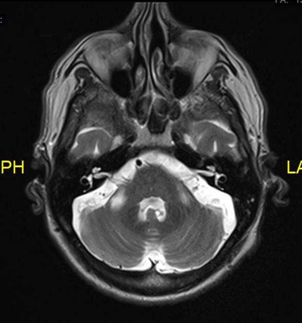
The majority of patients presenting with bilateral middle cerebellar peduncle lesions have high T2 signal without suppression on FLAIR, with matching low signal on T1 weighted images 4. This will usually have facilitated diffusion on ADC maps.
Diffusion restriction (low ADC values) may be seen acutely in infarction, lymphoma and acute cerebellitis 4,12. Enhancement is usually absent although some enhancement may be seen in lymphoma 4.
-
1. Okamoto K, Tokiguchi S, Furusawa T et-al. MR features of diseases involving bilateral middle cerebellar peduncles. AJNR Am J Neuroradiol. 24 (10): 1946-54. AJNR Am J Neuroradiol (full text) - Pubmed citation
-
2. Na SJ, Lee KO, Kim JE et-al. A case of cerebral erdheim-chester disease with progressive cerebellar syndrome. J Clin Neurol. 2008;4 (1): 45-50. doi:10.3988/jcn.2008.4.1.45 - Free text at pubmed - Pubmed citation
-
3. Uchino A, Sawada A, Takase Y, Kudo S. Symmetrical Lesions of the Middle Cerebellar Peduncle: MR Imaging and Differential Diagnosis. Magn Reson Med Sci. 2004;3(3):133-40. doi:10.2463/mrms.3.133 - Pubmed
-
4. Jiang J, Wang J, Lin M, Wang X, Zhao J, Shang X. Bilateral Middle Cerebellar Peduncle Lesions: Neuroimaging Features and Differential Diagnoses. Brain and Behavior. 2020;10(10):e01778. doi:10.1002/brb3.1778 - Pubmed
-
5. Morales H & Tomsick T. Middle Cerebellar Peduncles: Magnetic Resonance Imaging and Pathophysiologic Correlate. World J Radiol. 2015;7(12):438-47. doi:10.4329/wjr.v7.i12.438 - Pubmed
-
6. Kalus S, King J, Lui E et-al. Fragile X-associated tremor/ataxia syndrome: An under-recognised cause of tremor and ataxia. J Clin Neurosci. 2016;23: 162-4. doi:10.1016/j.jocn.2015.08.010 - Pubmed citation
-
7. Hiroaki Nozaki, Yumi Sekine, Toshio Fukutake, Yoshinori Nishimoto, Yutaka Shimoe, Akiko Shirata, Sohei Yanagawa, Mikio Hirayama, Masato Tamura, Masatoyo Nishizawa, Osamu Onodera. Characteristic features and progression of abnormalities on MRI for CARASIL. (2015) Neurology. 85 (5): 459. doi:10.1212/WNL.0000000000001803 - Pubmed
-
8. De Simone T, Regna-Gladin C, Carriero M, Farina L, Savoiardo M. Wallerian Degeneration of the Pontocerebellar Fibers. AJNR Am J Neuroradiol. 2005;26(5):1062-5. PMC8158597 - Pubmed
-
9. Guo Z, Lu T, Peng L et al. CLCN2-Related Leukoencephalopathy: A Case Report and Review of the Literature. BMC Neurol. 2019;19(1):156. doi:10.1186/s12883-019-1390-7 - Pubmed
-
10. Banks S, Morris P, Chen J et al. Brainstem and Cerebellar Involvement in MOG-IgG-Associated Disorder Versus Aquaporin-4-IgG and MS. J Neurol Neurosurg Psychiatry. 2020;92(4):384-90. doi:10.1136/jnnp-2020-325121 - Pubmed
-
11. Tang Y, Suddarth B, Du X, Matsumoto J. Reversible Diffusion Restriction of the Middle Cerebellar Peduncles and Dentate Nucleus in Acute Respiratory Syncytial Virus Cerebellitis: A Case Report. Emerg Radiol. 2013;21(1):89-92. doi:10.1007/s10140-013-1157-1 - Pubmed
-
12. Takanashi J, Miyamoto T, Ando N et al. Clinical and Radiological Features of Rotavirus Cerebellitis. AJNR Am J Neuroradiol. 2010;31(9):1591-5. doi:10.3174/ajnr.A2131 - Pubmed
Multiple choice questions:
Promoted articles (advertising)


 Unable to process the form. Check for errors and try again.