Cauda equina neuroendocrine tumour
- Biogen Australia Pty Ltd, Investigator-Initiated Research Grant for CAD software in multiple sclerosis: finished Oct 2021 (past)
Updates to Article Attributes
Spinal or cauda equina neuroendocrine tumours are rare sporadic WHO grade 1 tumours, almost invariably found below the conus arising either from the filum terminal or less commonly from the cauda equina 5.
Terminology
These tumours were previously known as spinal paragangliomas however they are molecularly and genetically distinct from paragangliomas seen elsewhere in the body and as such the term neuroendocrine tumour is preferred 5.
Epidemiology
The age range at presentation for spinal paragangliomas varies from childhood to older age (9-75 years of age) with most cases being diagnosed in middle age (30-60 years) 5. Males are somewhat more commonly affected than females 1-5.
Unlike paragangliomas/phaeochromocytomas that are commonly encountered as part of inherited syndromes, cauda equina neuroendocrine tumours are almost always sporadic 5.
Clinical presentation
The clinical presentation is either with local mass effect or neuroendocrine symptoms.
The most common presenting symptoms are lower back pain and sciatica from mass effect.
Cauda equina neuroendocrine tumour frequently actively secrete neuropeptides, particularly 5-hydroxytryptamine and somatostatin, although symptoms related to this chemical production are usually absent.
Additionally, these lesions may be a rare source of superficial siderosis, and thus present with sensory neural impairment, cranial nerve dysfunction and myelopathy 4.
Pathology
Paragangliomas of the spine usually arise in the region of the cauda equina, although rare reports of lesions elsewhere along the spine are available. They are indolent and considered WHO grade 1 tumours in the 5th edition (2021) WHO classification of CNS tumours 5.
Macroscopic appearance
Cauda equina neuroendocrine tumours are highly vascular masses, the majority (75%) of paragangliomas are encapsulated, usually attached to the filum terminale or less commonly a nerve root 5.
Microscopic appearance
All paragangliomas consist of two types of cells; type I and type II. The main components are lobules or nests of chief cells (type I); these structures are known as Zellballen. They are surrounded by a single layer of sustentacular cells (type II) 5.
Immunophenotype
Immunohistochemical examination confirms neuroendocrine differentiation of chief cells (type I) 5:
- chromogranin-A: positive
- synaptophysin: positive
Sustentacular cells (type II):
Genetics
Numerous mutations have been identified as predisposing to paragangliomas including VHL, RET, NF1, SDHAF2, SDHB, SDHC, SDHD, TNEM127 and MAX 5.
Radiographic features
Plain film
Plain films are unlikely to demonstrate any abnormality, although vertebral scalloping or rarely calcification may be seen 4.
Angiography
Conventional angiography demonstrates an intense early blush that persists into the late arterial and early venous phases.
CT
Spinal paragangliomas appear as soft tissue masses inferior to the conus. If contrast is administered they enhance vividly (as do paragangliomas elsewhere).
Large lesions may rarely demonstrate osseous erosion or remodelling of the adjacent vertebrae (vertebral scalloping). Rarely, these lesions calcify 4.
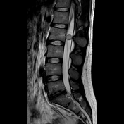
MRI
Similarly, on MRI these masses usually appear as well-circumscribed masses, inferior to the conus.
With some lesions, the characteristic salt-and-pepper appearance of neck and skull base paragangliomas may also be seen. Associated syringohydromyelia has been reported in some cases.
Reported signal characteristics include:
- T1: isointense
-
T2
- hyperintense
- flow voids are typically seen along the surface of and within the tumour nodule
- haemorrhage is common, leading to a haemosiderin cap sign
- T1 C+ (Gd): intense enhancement is virtually always seen
Treatment and prognosis
Surgical resection is the treatment of choice, sometimes with preoperative embolisation to reduce intra-operative blood loss. Resection alone is usually curative with a post-resection recurrence rate of less than 5% 1-5.
Rarely CSF dissemination is encountered 5.
Differential diagnosis
The differential diagnosis is essentially that of other intradural extramedullary tumours, particularly those located in the lumbar region.
- myxopapillary ependymoma
- neurogenic tumour
- spinal meningioma (rare in the lumbar region)
-<p><strong>Spinal </strong>or<strong> cauda equina neuroendocrine tumours</strong> are rare sporadic WHO grade 1 tumours, almost invariably found below the conus arising either from the filum terminal or less commonly from the cauda equina <sup>5</sup>. </p><h4>Terminology</h4><p>These tumours were previously known as <strong>spinal paragangliomas</strong> however they are molecularly and genetically distinct from <a href="/articles/paraganglioma-1">paragangliomas</a> seen elsewhere in the body and as such the term neuroendocrine tumour is preferred <sup>5</sup>.</p><h4>Epidemiology</h4><p>The age range at presentation for spinal paragangliomas varies from childhood to older age (9-75 years of age) with most cases being diagnosed in middle age (30-60 years) <sup>5</sup>. Males are somewhat more commonly affected than females <sup>1-5</sup>.</p><p>Unlike paragangliomas/phaeochromocytomas that are commonly encountered as part of inherited syndromes, cauda equina neuroendocrine tumours are almost always sporadic <sup>5</sup>. </p><h4>Clinical presentation</h4><p>The clinical presentation is either with local mass effect or neuroendocrine symptoms. </p><p>The most common presenting symptoms are lower back pain and sciatica from mass effect.</p><p>Cauda equina neuroendocrine tumour frequently actively secrete neuropeptides, particularly 5-hydroxytryptamine and somatostatin, although symptoms related to this chemical production are usually absent. </p><p>Additionally, these lesions may be a rare source of <a href="/articles/superficial-siderosis-1">superficial siderosis</a>, and thus present with sensory neural impairment, cranial nerve dysfunction and myelopathy <sup>4</sup>.</p><h4>Pathology</h4><p>Paragangliomas of the spine usually arise in the region of the cauda equina, although rare reports of lesions elsewhere along the spine are available. They are indolent and considered WHO grade 1 tumours in the 5th edition (2021) <a href="/articles/who-classification-of-cns-tumours-1">WHO classification of CNS tumours</a> <sup>5</sup>. </p><h5>Macroscopic appearance</h5><p>Cauda equina neuroendocrine tumours are highly vascular masses, the majority (75%) of paragangliomas are encapsulated, usually attached to the filum terminale or less commonly a nerve root <sup>5</sup>. </p><h5>Microscopic appearance</h5><p>All paragangliomas consist of two types of cells; type I and type II. The main components are lobules or nests of chief cells (type I); these structures are known as Zellballen. They are surrounded by a single layer of sustentacular cells (type II) <sup>5</sup>.</p><h5>Immunophenotype</h5><p>Immunohistochemical examination confirms neuroendocrine differentiation of chief cells (type I) <sup>5</sup>: </p><ul>-<li>-<a href="/articles/chromogranin-a">chromogranin-A</a>: positive</li>-<li>-<a href="/articles/synaptophysin">synaptophysin</a>: positive</li>-</ul><p>Sustentacular cells (type II):</p><ul>-<li>-<a href="/articles/s100">S100</a>: usually positive</li>-<li>-<a href="/articles/glial-fibrillary-acid-protein-gfap">GFAP</a>: usually positive</li>-</ul><h5>Genetics</h5><p>Numerous mutations have been identified as predisposing to paragangliomas including <em>VHL</em>, <em>RET</em>, <em>NF1</em>, <em>SDHAF2</em>, <em>SDHB</em>, <em>SDHC</em>, <em>SDHD</em>, <em>TNEM127</em> and <em>MAX</em> <sup>5</sup>. </p><h4>Radiographic features</h4><h5>Plain film</h5><p>Plain films are unlikely to demonstrate any abnormality, although <a href="/articles/vertebral-scalloping">vertebral scalloping</a> or rarely calcification may be seen <sup>4</sup>. </p><h5>Angiography</h5><p>Conventional angiography demonstrates an intense early blush that persists into the late arterial and early venous phases.</p><h5>CT</h5><p>Spinal paragangliomas appear as soft tissue masses inferior to the conus. If contrast is administered they enhance vividly (as do paragangliomas elsewhere). </p><p>Large lesions may rarely demonstrate osseous erosion or remodelling of the adjacent vertebrae (<a href="/articles/vertebral-scalloping">vertebral scalloping</a>). Rarely, these lesions calcify <sup>4</sup>. </p><h5>MRI</h5><p>Similarly, on MRI these masses usually appear as well-circumscribed masses, inferior to the conus. </p><p>With some lesions, the characteristic <a href="/articles/salt-and-pepper-sign-disambiguation">salt-and-pepper</a> appearance of neck and skull base paragangliomas may also be seen. Associated <a href="/articles/syrinx-1">syringohydromyelia</a> has been reported in some cases.</p><p>Reported signal characteristics include:</p><ul>-<li>-<strong>T1:</strong> isointense</li>-<li>-<strong>T2</strong><ul>-<li>hyperintense</li>-<li>flow voids are typically seen along the surface of and within the tumour nodule</li>-<li>haemorrhage is common, leading to a <a href="/articles/cap-sign">haemosiderin cap sign</a>-</li>-</ul>-</li>-<li>-<strong>T1 C+ (Gd):</strong> intense enhancement is virtually always seen</li>-</ul><h4>Treatment and prognosis</h4><p>Surgical resection is the treatment of choice, sometimes with preoperative embolisation to reduce intra-operative blood loss. Resection alone is usually curative with a post-resection recurrence rate of less than 5% <sup>1-5</sup>.</p><p>Rarely CSF dissemination is encountered <sup>5</sup>. </p><h4>Differential diagnosis</h4><p>The differential diagnosis is essentially that of other <a href="/articles/intradural-extramedullary-spinal-tumours-1">intradural extramedullary tumours</a>, particularly those located in the lumbar region. </p><ul>-<li><a href="/articles/myxopapillary-ependymoma-1">myxopapillary ependymoma</a></li>-<li>-<a href="/articles/neurogenic-tumours-1">neurogenic tumour</a><ul>-<li><a href="/articles/spinal-schwannoma">spinal schwannoma</a></li>-<li><a href="/articles/spinal-neurofibroma">spinal neurofibroma</a></li>-</ul>-</li>-<li>-<a href="/articles/spinal-meningioma">spinal meningioma</a> (rare in the lumbar region)</li>- +<p><strong>Spinal </strong>or<strong> cauda equina neuroendocrine tumours</strong> are rare sporadic WHO grade 1 tumours, almost invariably found below the conus arising either from the filum terminal or less commonly from the cauda equina <sup>5</sup>. </p><h4>Terminology</h4><p>These tumours were previously known as <strong>spinal paragangliomas</strong> however they are molecularly and genetically distinct from <a href="/articles/paraganglioma-1">paragangliomas</a> seen elsewhere in the body and as such the term neuroendocrine tumour is preferred <sup>5</sup>.</p><h4>Epidemiology</h4><p>The age range at presentation for spinal paragangliomas varies from childhood to older age (9-75 years of age) with most cases being diagnosed in middle age (30-60 years) <sup>5</sup>. Males are somewhat more commonly affected than females <sup>1-5</sup>.</p><p>Unlike paragangliomas/phaeochromocytomas that are commonly encountered as part of inherited syndromes, cauda equina neuroendocrine tumours are almost always sporadic <sup>5</sup>. </p><h4>Clinical presentation</h4><p>The clinical presentation is either with local mass effect or neuroendocrine symptoms. </p><p>The most common presenting symptoms are lower back pain and sciatica from mass effect.</p><p>Cauda equina neuroendocrine tumour frequently actively secrete neuropeptides, particularly 5-hydroxytryptamine and somatostatin, although symptoms related to this chemical production are usually absent. </p><p>Additionally, these lesions may be a rare source of <a href="/articles/superficial-siderosis-1">superficial siderosis</a>, and thus present with sensory neural impairment, cranial nerve dysfunction and myelopathy <sup>4</sup>.</p><h4>Pathology</h4><p>Paragangliomas of the spine usually arise in the region of the cauda equina, although rare reports of lesions elsewhere along the spine are available. They are indolent and considered WHO grade 1 tumours in the 5th edition (2021) <a href="/articles/who-classification-of-cns-tumours-1">WHO classification of CNS tumours</a> <sup>5</sup>. </p><h5>Macroscopic appearance</h5><p>Cauda equina neuroendocrine tumours are highly vascular masses, the majority (75%) of paragangliomas are encapsulated, usually attached to the filum terminale or less commonly a nerve root <sup>5</sup>. </p><h5>Microscopic appearance</h5><p>All paragangliomas consist of two types of cells; type I and type II. The main components are lobules or nests of chief cells (type I); these structures are known as Zellballen. They are surrounded by a single layer of sustentacular cells (type II) <sup>5</sup>.</p><h5>Immunophenotype</h5><p>Immunohistochemical examination confirms neuroendocrine differentiation of chief cells (type I) <sup>5</sup>: </p><ul>
- +<li>
- +<a href="/articles/chromogranin-a">chromogranin-A</a>: positive</li>
- +<li>
- +<a href="/articles/synaptophysin">synaptophysin</a>: positive</li>
- +</ul><p>Sustentacular cells (type II):</p><ul>
- +<li>
- +<a href="/articles/s100">S100</a>: usually positive</li>
- +<li>
- +<a href="/articles/glial-fibrillary-acid-protein-gfap">GFAP</a>: usually positive</li>
- +</ul><h5>Genetics</h5><p>Numerous mutations have been identified as predisposing to paragangliomas including <em>VHL</em>, <em>RET</em>, <em>NF1</em>, <em>SDHAF2</em>, <em>SDHB</em>, <em>SDHC</em>, <em>SDHD</em>, <em>TNEM127</em> and <em>MAX</em> <sup>5</sup>. </p><h4>Radiographic features</h4><h5>Plain film</h5><p>Plain films are unlikely to demonstrate any abnormality, although <a href="/articles/vertebral-scalloping">vertebral scalloping</a> or rarely calcification may be seen <sup>4</sup>. </p><h5>Angiography</h5><p>Conventional angiography demonstrates an intense early blush that persists into the late arterial and early venous phases.</p><h5>CT</h5><p>Spinal paragangliomas appear as soft tissue masses inferior to the conus. If contrast is administered they enhance vividly (as do paragangliomas elsewhere). </p><p>Large lesions may rarely demonstrate osseous erosion or remodelling of the adjacent vertebrae (<a href="/articles/vertebral-scalloping">vertebral scalloping</a>). Rarely, these lesions calcify <sup>4</sup>. </p><h5>MRI</h5><p>Similarly, on MRI these masses usually appear as well-circumscribed masses, inferior to the conus. </p><p>With some lesions, the characteristic <a href="/articles/salt-and-pepper-sign-disambiguation">salt-and-pepper</a> appearance of neck and skull base paragangliomas may also be seen. Associated <a href="/articles/syrinx-1">syringohydromyelia</a> has been reported in some cases.</p><p>Reported signal characteristics include:</p><ul>
- +<li>
- +<strong>T1:</strong> isointense</li>
- +<li>
- +<strong>T2</strong><ul>
- +<li>hyperintense</li>
- +<li>flow voids are typically seen along the surface of and within the tumour nodule</li>
- +<li>haemorrhage is common, leading to a <a href="/articles/cap-sign">haemosiderin cap sign</a>
- +</li>
- +</ul>
- +</li>
- +<li>
- +<strong>T1 C+ (Gd):</strong> intense enhancement is virtually always seen</li>
- +</ul><h4>Treatment and prognosis</h4><p>Surgical resection is the treatment of choice, sometimes with preoperative embolisation to reduce intra-operative blood loss. Resection alone is usually curative with a post-resection recurrence rate of less than 5% <sup>1-5</sup>.</p><p>Rarely CSF dissemination is encountered <sup>5</sup>. </p><h4>Differential diagnosis</h4><p>The differential diagnosis is essentially that of other <a href="/articles/intradural-extramedullary-spinal-tumours-1">intradural extramedullary tumours</a>, particularly those located in the lumbar region. </p><ul>
- +<li><a href="/articles/myxopapillary-ependymoma-1">myxopapillary ependymoma</a></li>
- +<li>
- +<a href="/articles/neurogenic-tumours-1">neurogenic tumour</a><ul>
- +<li><a href="/articles/spinal-schwannoma">spinal schwannoma</a></li>
- +<li><a href="/articles/spinal-neurofibroma">spinal neurofibroma</a></li>
- +</ul>
- +</li>
- +<li>
- +<a href="/articles/spinal-meningioma">spinal meningioma</a> (rare in the lumbar region)</li>
Image 6 MRI (T2) ( create )