Citation, DOI, disclosures and article data
Citation:
Ibrahim D, Weerakkody Y, Baba Y, et al. Chronic otomastoiditis with tympanosclerosis. Reference article, Radiopaedia.org (Accessed on 20 Feb 2025) https://doi.org/10.53347/rID-47520
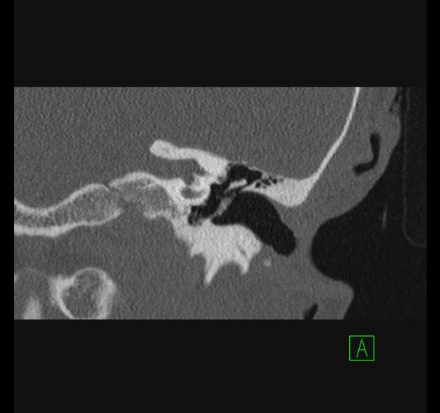
Chronic otomastoiditis with tympanosclerosis represents calcific foci within the middle ear or tympanic membrane secondary to suppurative chronic otomastoiditis.
Tympanosclerosis reflects deposits of hyalinized collagen in the tympanic cavity. It can manifest as unifocal or multifocal punctate or web-like calcifications in the middle ear cavity or on the tympanic membrane (myringosclerosis).
Features include chronic otomastoiditis findings such as middle ear soft tissue density and under-pneumatized mastoid associated with calcifications. Common locations of calcifications include:
-
1. Harnsberger HR, MBBS CMG, Michel MA et-al. Diagnostic Imaging Head and Neck. Lippincott Williams & Wilkins. ISBN:1931884781. Read it at Google Books - Find it at Amazon
-
2. Kadam PD, Chuan HH. Erratum to: Rectocutaneous fistula with transmigration of the suture: a rare delayed complication of vault fixation with the sacrospinous ligament. Int Urogynecol J. 2016;27 (3): 505. doi:10.1007/174_2012_788 - Pubmed citation
-
3. Larem A, Abu Rajab Altamimi Z, Aljariri A et al. Reliability of high‐resolution CT Scan in Diagnosis of Ossicular Tympanosclerosis. Laryngoscope Investigative Otolaryngology. 2021;6(3):540-8. doi:10.1002/lio2.594 - Pubmed
Promoted articles (advertising)


 Unable to process the form. Check for errors and try again.