
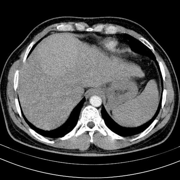
Differential diagnoses of cirrhotic liver nodules include regenerative liver nodules, dysplastic liver nodules, and hepatocellular carcinoma (HCC), all represent a spectrum of diseases ranging from non-neoplastic reparative process (regenerative) to nuclear atypia (dysplastic) to typical neoplasia (HCC). It's not always possible to differentiate the three types of lesion on imaging, however, there are some suggestive features that may favour either one. These features are summarised in this article.
On this page:
Regenerative liver nodules
- numerous (thousands) micro (< 3 mm) nodules, but can be single/few macro (> 3 mm) or giant (> 5 mm) nodules
- invisible on non-contrast CT, except for siderotic nodules which appear hyperdense
- siderotic nodules show blooming artifact on T2*
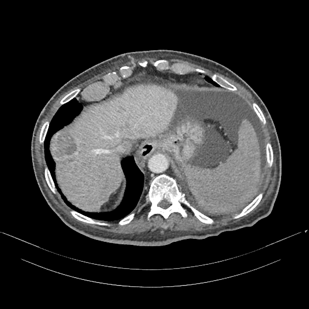
- do not enhance, or enhances similar to or lower than the liver parenchyma
- no arterial hyperenhancement
Dysplastic liver nodule
- single, or few nodules
- can be hypo-, iso-, or hyperdense on CT
- early arterial enhancement without washout on later phases
- iso- to hypointense on T2WI
- no restricted diffusion
Early hepatocellular carcinoma
- often hypodense on CT
- early arterial enhancement with washout on later phases
- slightly hyperintense on T2WI
- may show restricted diffusion


 Unable to process the form. Check for errors and try again.