
CT of the adrenal glands is a study utilised in patients with incidentally discovered adrenal lesions on other studies, in order to characterise the lesions, and to seek adrenal abnormalities in patients with hormonal biochemical abnormalities.
On this page:
Indications
Characterise incidentally discovered adrenal nodules and seek adrenal abnormalities when clinically suspected.
Purpose
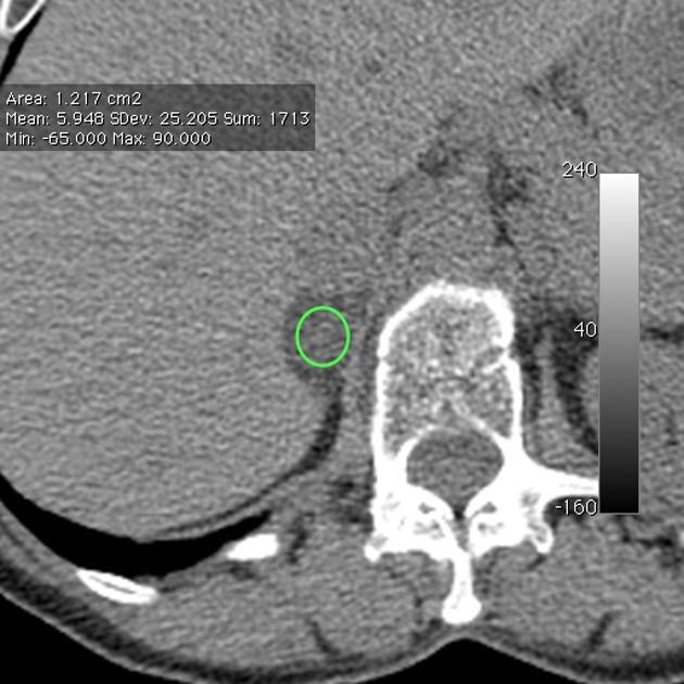
To characterise adrenal nodules on the basis of the density on non-contrast and post-contrast imaging, and washout characteristics. Nodules with lipid density measurements on the non-contrast series can be diagnosed as lipid-rich adenomas, and in nodules that are not lipid-rich, washout calculations can be used to differentiate between lipid-poor adenomas and indeterminate lesions, with differentials for the latter including adrenocortical carcinoma and phaeochromocytoma.
Technique
-
patient position
supine with their arms above their head
-
scout
diaphragm to iliac crests
-
scan extent
diaphragm to iliac crests
-
scan direction
craniocaudal
-
scan delay
non-contrast series: no delay
portal venous phase series: 60-70 second delay
delayed phase series: 15-minute delay
-
respiration phase
inspiration, breath-hold
Practical points
the choice of which incidental lesion to investigate may be determined by published algorithms such as the ACR White Paper
density measurements <10 HU using region-of-interest calculations on a non-contrast series indicates a lipid-rich adenoma or a myelolipoma
if the density is >10 HU on the non-contrast series, the density measurements on the portal venous and delayed phase studies may be used to calculate the relative and absolute washout
External links
If any of these links are broken or for other problems and questions, please contact editors@radiopaedia.org.


 Unable to process the form. Check for errors and try again.