
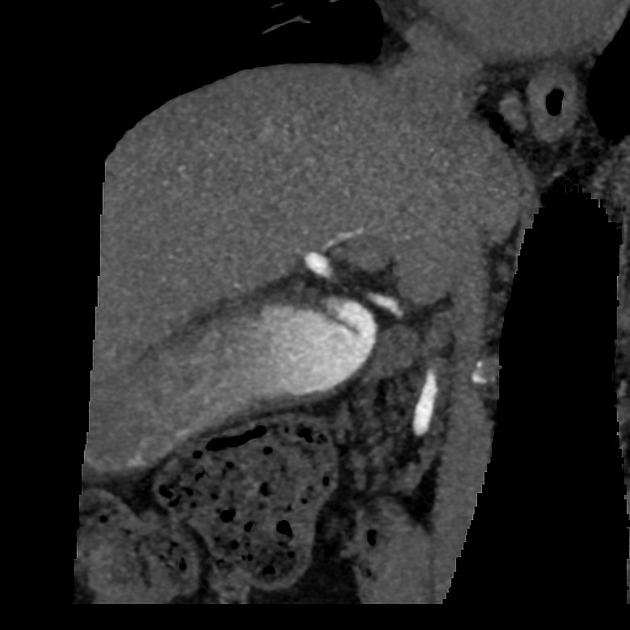
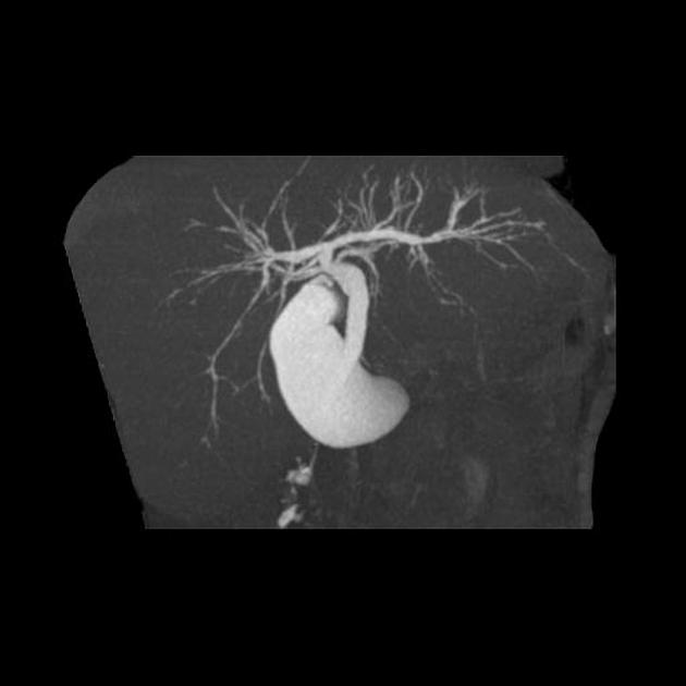
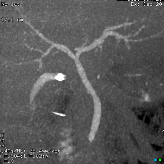
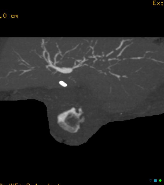
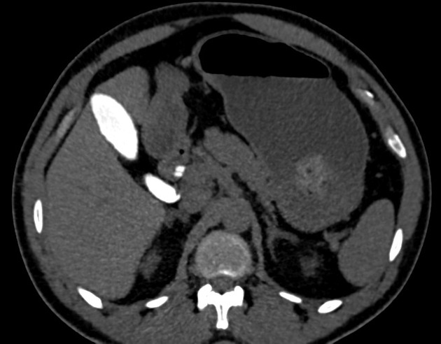
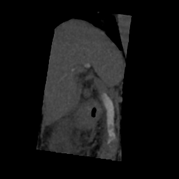
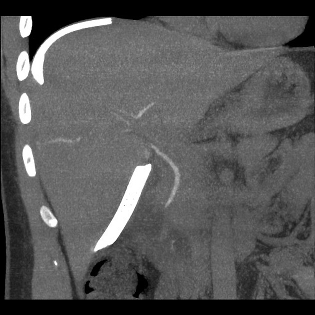
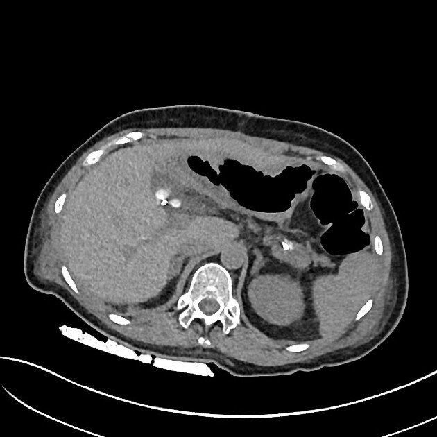
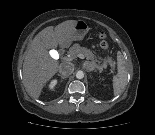
CT cholangiography is a technique of imaging the biliary tree with the usage of hepatobiliary excreted contrast. It is useful in delineating biliary anatomy, identifying a bile leak or looking for retained gallstones within the biliary system.
On this page:
Indications
Second-line test (after ultrasound) when investigating for right upper quadrant pain, obstructive LFTs, etc. It can also be used in the postoperative setting (e.g. post-cholecystectomy) where there is a concern for common bile duct injury or retained gallstones, or where intraoperative cholangiography (IOC) is unable to be performed due to extensive inflammation or a narrow cystic duct, proving direct cannulation difficult.
Purpose
The purpose of CT cholangiography is to identify a filling defect in the biliary tree that represents choledocholithiasis or a contrast leak from the biliary tree in case of injury.
Contraindications
bilirubin should be <30 µmol/L nor should it be rising rapidly (as the impaired excretory ability of hepatocytes can affect contrast excretion in bile)
severe hepatic or renal dysfunction
thyroid dysfunction
Technique
CT cholangiography may be performed with either intravenous or oral cholangiographic contrast agents both of which outline the biliary tree with positive contrast.
Agents
meglumine iotroxate (Biliscopin): intravenous CT cholangiography agent
Findings
bile leak / biloma
aberrant biliary tree anatomy
other causes of biliary tree obstruction, e.g. pancreatic head tumors
Alternative examinations
contrast-enhanced MR cholangiography: with gadolinium ethoxybenzyl diethylenetriamine pentaacetic acid
endoscopic retrograde cholangiopancreatography (ERCP): carries a 5% risk of post-procedure pancreatitis
intraoperative cholangiography (IOC): performed during cholecystectomy to allow intraoperative detection of retained gallstones or common bile duct injury
Mimics
occasionally vicarious contrast material excretion can give opacification of the gallbladder and biliary system


 Unable to process the form. Check for errors and try again.