
Deep cervical fascia
Citation, DOI, disclosures and article data
At the time the article was created Craig Hacking had no recorded disclosures.
View Craig Hacking's current disclosuresAt the time the article was last revised Tee Yu Jin had no recorded disclosures.
View Tee Yu Jin's current disclosures- cervical fascia
The deep cervical fascia consists of three separate but related fascial layers that encircle structures in the neck and allow anatomic compartmentalisation into the deep spaces of the head and neck. Each layer contributes to the carotid sheath. See the separate articles for further details:
- superficial layer of the deep cervical fascia
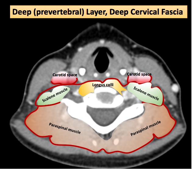
- middle layer of the deep cervical fascia
- deep layer of the deep cervical fascia
On this page:
Terminology
The deep cervical fascia was historically defined in contrast to the superficial cervical fascia, the latter of which primarily includes the platysma and subcutaneous fat and vessels. However, as with other fascia in the body, use of the terminology of the superficial cervical fascia has declined in favor of "subcutaneous tissue" 4. Thus, an unspecified reference to cervical fascia mainly refers to the deep cervical fascia.
Radiographic features
The fascia usually cannot be visualized directly by imaging.
ADVERTISEMENT: Supporters see fewer/no ads
Related pathology
The fascial layers limit the spread of disease. Knowledge of fascial anatomy helps identify common routes of spread of infection and metastatic disease in the head and neck, including spread into the chest.
The fascial layers also provide surgical cleavage planes.
See also
References
- 1. Last's anatomy, regional and applied. Churchill Livingstone. ISBN:044304662X. Read it at Google Books - Find it at Amazon
- 2. Moore KL, Agur AMR, Dalley AF. Clinically oriented anatomy. LWW. ISBN:1451119453. Read it at Google Books - Find it at Amazon
- 3. Butler, P, 1999. Applied Radiological Anaomy. 1st ed. London: Cambridge University Press.
- 4. Guidera AK, Dawes PJ, Fong A, Stringer MD. Head and neck fascia and compartments: no space for spaces. (2014) Head & neck. 36 (7): 1058-68. doi:10.1002/hed.23442 - Pubmed
- 5. Peter M. Som, Hugh D. Curtin. Head and Neck Imaging - 2 Volume Set. (2019) ISBN: 9780323053556
- 6. Kamalian S, Avery L, Lev M, Schaefer P, Curtin H, Kamalian S. Nontraumatic Head and Neck Emergencies. (2019) RadioGraphics. 39 (6): 1808-1823. doi:10.1148/rg.2019190159 - Pubmed
Incoming Links
- Thymus
- Thoracic plane
- Parapharyngeal space
- Punctum nervosum
- Peritonsillar space
- Bezold abscess
- Common carotid artery
- Perivertebral space
- Brachial plexus
- Superficial cervical fascia
- Submandibular gland
- Trapezius muscle
- Transverse cervical nerve
- Thyrocervical trunk
- Stylomandibular ligament
- Digastric muscle
- Superficial layer of the deep cervical fascia
- Thyroid gland
- Sialadenitis
- Supraclavicular triangle
Related articles: Anatomy: Head and neck
- skeleton of the head and neck
-
cranial vault
- scalp (mnemonic)
- fontanelle
-
sutures
- calvarial
- facial
- frontozygomatic suture
- frontomaxillary suture
- frontolacrimal suture
- frontonasal suture
- temporozygomatic suture
- zygomaticomaxillary suture
- parietotemporal suture (parietomastoid suture)
- occipitotemporal suture (occipitomastoid suture)
- sphenofrontal suture
- sphenozygomatic suture
- spheno-occipital suture (not a true suture)
- lacrimomaxillary suture
- nasomaxillary suture
- internasal suture
- basal/internal
- skull landmarks
- frontal bone
- temporal bone
- parietal bone
- occipital bone
- skull base (foramina)
-
facial bones
- midline single bones
- paired bilateral bones
- cervical spine
- hyoid bone
- laryngeal cartilages
-
cranial vault
- muscles of the head and neck
- muscles of the tongue (mnemonic)
- muscles of mastication
-
facial muscles
- epicranius muscle
- circumorbital and palpebral muscles
- nasal muscles
-
buccolabial muscles
- elevators, retractors and evertors of the upper lip
- levator labii superioris alaeque nasalis muscle
- levator labii superioris muscle
- zygomaticus major muscle
- zygomaticus minor muscle
- levator anguli oris muscle
- malaris muscle
- risorius muscle
- depressors, retractors and evertors of the lower lip
- depressor labii inferioris muscle
- depressor anguli oris muscle
- mentalis muscle
- compound sphincter
-
orbicularis oris muscle
- incisivus labii superioris muscle
- incisivus labii inferioris muscle
-
orbicularis oris muscle
- muscle of mastication
- modiolus
- elevators, retractors and evertors of the upper lip
- muscles of the middle ear
- orbital muscles
- muscles of the soft palate
- pharyngeal muscles
- suprahyoid muscles
- infrahyoid muscles
- intrinsic muscles of the larynx
- muscles of the neck
- platysma muscle
- longus colli muscle
- longus capitis muscle
- scalenus anterior muscle
- scalenus medius muscle
- scalenus posterior muscle
- scalenus pleuralis muscle
- sternocleidomastoid muscle
-
suboccipital muscles
- rectus capitis posterior major muscle
- rectus capitis posterior minor muscle
- obliquus capitis superior muscle
- obliquus capitis inferior muscle
- accessory muscles of the neck
- deep cervical fascia
-
deep spaces of the neck
- anterior cervical space
- buccal space
- carotid space
- danger space
- deep cervical fascia
- infratemporal fossa
- masticator space
- parapharyngeal space
- stylomandibular tunnel
- parotid space
- pharyngeal (superficial) mucosal space
- perivertebral space
- posterior cervical space
- pterygopalatine fossa
- retropharyngeal space
- suprasternal space (of Burns)
- visceral space
- surgical triangles of the neck
- orbit
- ear
- paranasal sinuses
- upper respiratory tract
- viscera of the neck
- blood supply of the head and neck
-
arterial supply
-
common carotid artery
- carotid body
- carotid bifurcation
- subclavian artery
- variants
-
common carotid artery
- venous drainage
-
arterial supply
- innervation of the head and neck
-
cranial nerves
- olfactory nerve (CN I)
- optic nerve (CN II)
- oculomotor nerve (CN III)
- trochlear nerve (CN IV)
-
trigeminal nerve (CN V) (mnemonic)
- trigeminal ganglion
- ophthalmic division
- maxillary division
- mandibular division
- abducens nerve (CN VI)
- facial nerve (CN VII)
-
vestibulocochlear nerve (CN VIII)
- vestibular ganglion (Scarpa's ganglion)
- glossopharyngeal nerve (CN IX)
- vagus nerve (CN X)
- (spinal) accessory nerve (CN XI)
- hypoglossal nerve (CN XII)
- parasympathetic ganglia of the head and neck
- cervical sympathetic ganglia
- greater occipital nerve
- third occipital nerve
-
cervical plexus
- muscular branches
- longus capitis
- longus colli
- scalenes
- geniohyoid
- thyrohyoid
-
ansa cervicalis
- omohyoid (superior and inferior bellies separately)
- sternothyroid
- sternohyoid
- phrenic nerve
- contribution to the accessory nerve (CN XI)
- cutaneous branches
- muscular branches
- brachial plexus
- pharyngeal plexus
-
cranial nerves
- lymphatic drainage of the head and neck
- embryological development of the head and neck


 Unable to process the form. Check for errors and try again.