
This is a basic article for medical students and other non-radiologists
Distal radial fractures are a relatively common group of injuries that usually occur following a fall. The commonest of these fractures is a transverse extra-articular fracture and where there is associated dorsal angulation, this is termed a Colles fracture.
On this page:
Reference article
This is a summary article. For more information, you can read a more in-depth reference articles: distal radial fracture, Colles fracture.
Summary
-
anatomy
- normal radius
-
epidemiology
- bimodal age and sex distribution
- younger males in high energy mechanisms
- older females after simple falls
-
presentation
- fall onto an outstretched hand with pain and deformity
-
pathophysiology
- after FOOSH force transmitted through the wrist
- a direct blow to the wrist may also result in a fracture
-
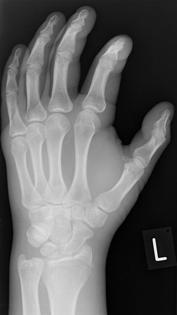
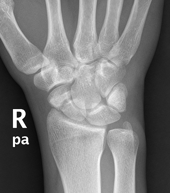
investigation
- wrist series (AP and lateral)
-
treatment
- often treatment is conservative with immobilisation in a cast
- if there is deformity and fracture angulation reduction is required
- in some cases, internal fixation is needed
Radiographic features
Plain radiograph
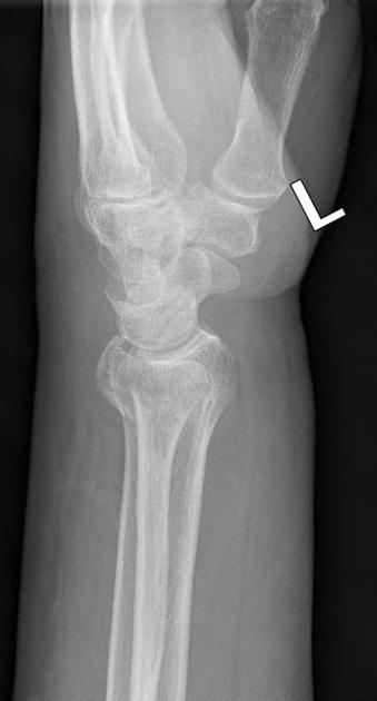
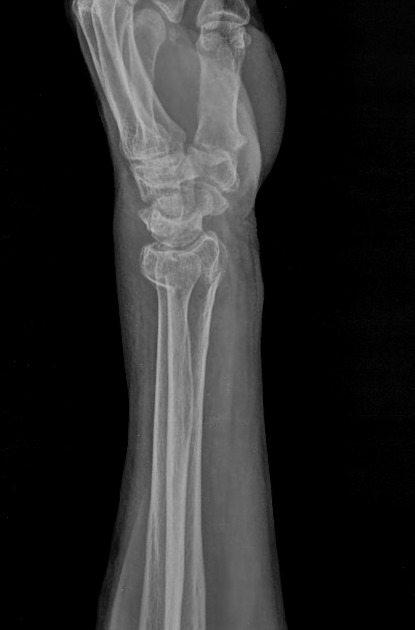
The commonest fracture of the distal radius is a transverse extra-articular fracture which is usually seen as a transverse lucency across the distal radius in the region of the metaphysis.
If there is impaction, the fracture may be seen as a sclerotic line.
Transverse fractures may be angulated - dorsal angulation is commonest (a Colles fracture). There may be fracture extension into the joint which is important to pick up.


 Unable to process the form. Check for errors and try again.