
The double aorta artifact is a relatively common ultrasound artifact, which can appear both on standard B-mode and color Doppler imaging, resulting in an artifactually duplicated abdominal aorta in the transverse plane. Knowledge of this artifact is paramount as potential differential diagnoses include life threatening conditions (e.g. aortic dissection).
On this page:
Physics
The artifact is caused by the differential refraction of the ultrasound beam while passing through the relatively echogenic rectus abdominis muscles, and the comparatively hypoechoic fatty tissue underneath. The interface between these tissues creates an acoustic prism effect. The artifact primarily affects curvilinear transducers 1.
Radiographic features
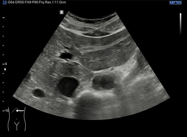
Partially, or less commonly completely duplicated, "double barrel" appearance of the abdominal aorta in the transverse plane is characteristic, which also affects color Doppler imaging. Adjacent structures (e.g. anterior vertebral echo) are duplicated too, but this is usually harder to appreciate.
Differential diagnosis
- abdominal aortic dissection or aneurysm
- congenital duplication of the aorta (exceedingly rare)
Practical points
The artifact can be diminished by changing the angle of insonation in a way that the beam no longer passes through the "acoustic prism" formed by the rectus abdominis muscles and the fatty tissue beneath the linea alba. Rotating the probe into the sagittal plane has a similar result. Note that pulsed wave Doppler ultrasound curves are not affected by this artifact, and will display normal spectral waveform, which also differentiates this phenomenon from its mimickers.
The artifact is more common in younger, athletic patients, and the duplication is usually incomplete 1.
Note that this phenomenon (albeit rarely) can be encountered in other regions, e.g. during echocardiography 2.


 Unable to process the form. Check for errors and try again.