
This is a basic article for medical students and other non-radiologists
Extra-axial collections are collections of fluid within the skull, but outside the brain parenchyma. They may be comprised of CSF, blood or pus and may exist in the extradural, subdural or subarachnoid space.
On this page:
Reference article
This is a summary article; we do not have a more in-depth reference article.
Summary
- anatomy
-
pathophysiology
- fluid collection in the extradural, subdural or subarachnoid space
- blood, e.g. post-traumatic extradural haemorrhage
- fluid, e.g. chronic subdural haemorrhage
- pus, e.g. subdural empyema
- fluid collection in the extradural, subdural or subarachnoid space
Imaging
-
role of imaging
- determine whether there is an extra-axial collection
- determine where the collection is:
-
subarachnoid
- extends down into the sulci
- extends into the basal cisterns
-
subdural
- more crescent-like shape
- may cross sutures
-
extradural
- lentiform (lens-like) shape
- do not cross cranial sutures
-
subarachnoid
- determine whether there is mass effect and:
-
radiographic features
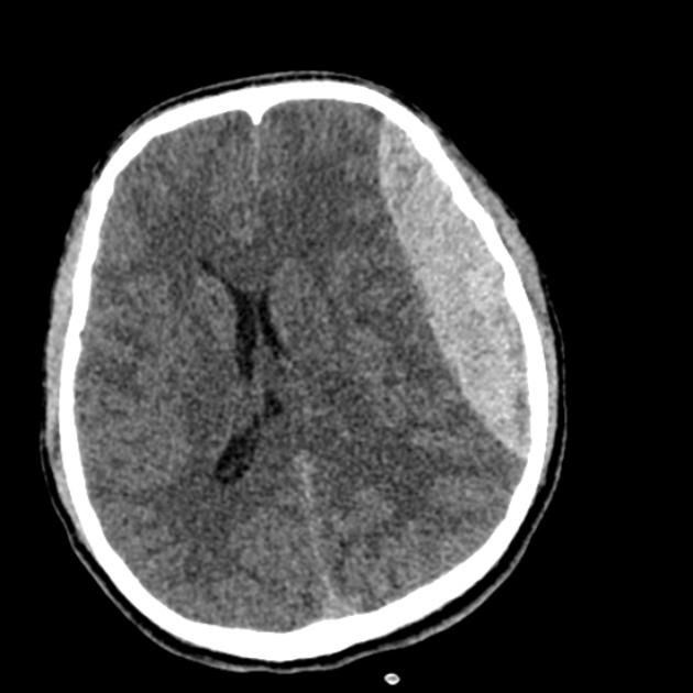
- CT head
- best initial test, especially in an unwell patient
- haemorrhage in the acute setting is white
- as blood ages, it becomes darker (more watery) on CT
- extra-axial collections are not always blood, e.g. empyema
- MRI head
- may be helpful for further assessment
- may be easier to determine which space the collection is in
- using different sequences, it should be possible to determine the type of fluid and narrow the differential diagnosis
- CT head


 Unable to process the form. Check for errors and try again.