
This is a basic article for medical students and other non-radiologists
Extradural haemorrhages (EDH) represent collections of blood in the extradural (epidural) space. The haemorrhage sits between the skull superficially and the dura which overlies the brain parenchyma.
The bleed in relation to the dura mater is the key anatomical difference between an extradural and a subdural haemorrhage. As a student, a helpful tip is to remember that the dura tightly adheres to the intracranial bony sutures. Thus, an extradural haemorrhage is confined within that space, often producing a biconvex (also called lentiform) shape on CT.
On this page:
Reference article
This is a summary article; read more in our article on extradural haemorrhage.
Summary
- anatomy
-
epidemiology
- young patients
- high-energy impact trauma
-
presentation
- headache (may be due to associated fracture)
- localising signs secondary to mass effect
- loss of consciousness
-
pathophysiology
- source of haemorrhage tends to be arterial
- associated skull fracture
- middle meningeal artery is particularly at risk
-
investigation
- CT head (non-contrast) is quick, easy and readily available
-
treatment
- urgent consideration of neurosurgical intervention
- surgical treatment: evacuation of clot through a burr hole
- smaller bleeds may be managed conservatively
Imaging
-
role of imaging
- initial diagnosis
- determine of associated injuries and sequela
- determine the underlying cause
- determine further imaging and follow up
-
radiographic features
- CT
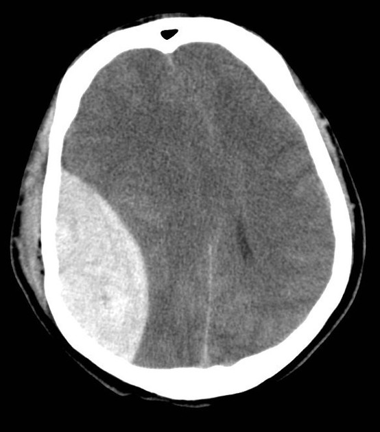
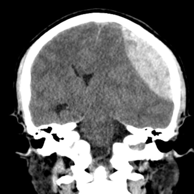
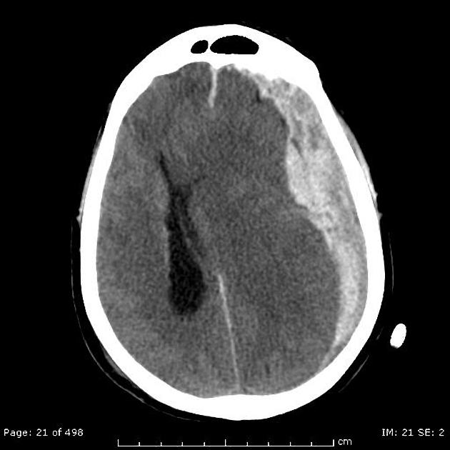
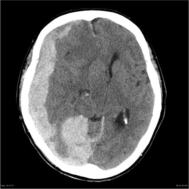
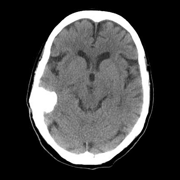
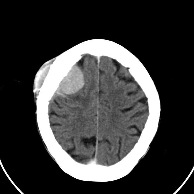
- peripheral hyperdense biconvex extra-axial collection
- looking like a lens (lentiform) or egg ("eggs"-tradural)
- well-demarcated haemorrhage between brain and skull
- acute: hyperdense
- active bleeding: central dark areas
- bound by skull sutures
- associated injuries, e.g. fracture, parenchymal contusion
- sequelae, e.g. mass effect and midline shift
- peripheral hyperdense biconvex extra-axial collection
- CT


 Unable to process the form. Check for errors and try again.