
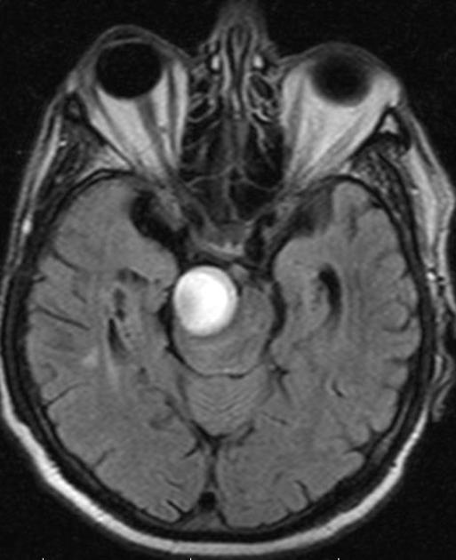
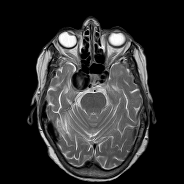
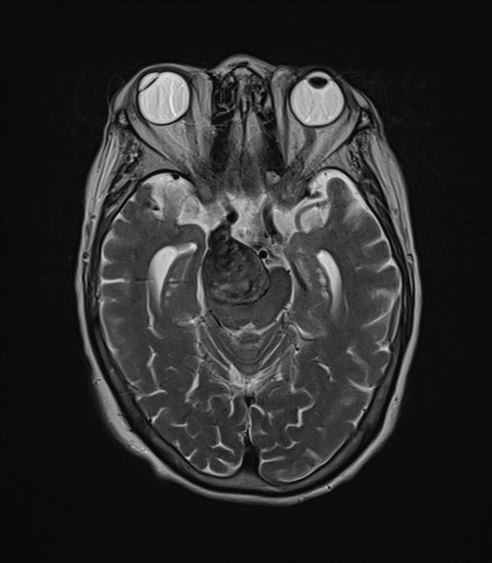
Giant cerebral aneurysms are ones that measure >25 mm in their greatest dimension.
On this page:
Epidemiology
Giant cerebral aneurysms account for ~5% of all intracranial aneurysms 1,3. They occur in the 5th-7th decades and are more common in females 2.
Clinical presentation
Patients can present with symptoms and signs of mass effect or subarachnoid haemorrhage 1,2.
Pathology
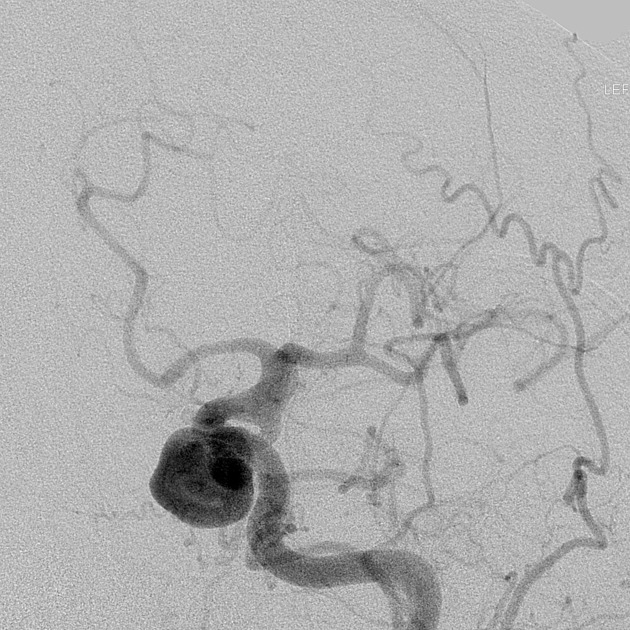
Most commonly represent saccular cerebral aneurysms but may also be fusiform or serpentine in morphology 1. They are thought to develop via two pathways 2:
internal elastic lamina de novo defect
enlargement from a smaller aneurysm
Location
Compared to non-giant cerebral aneurysms there is an increased incidence in the posterior circulation (~35%) 3.
Radiographic features
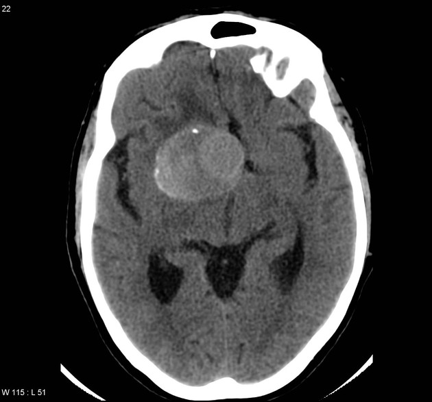
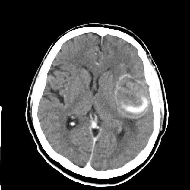
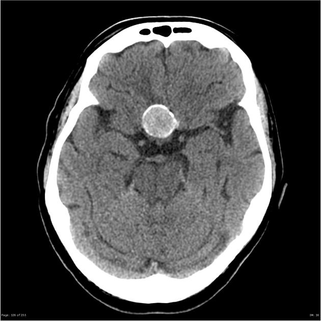
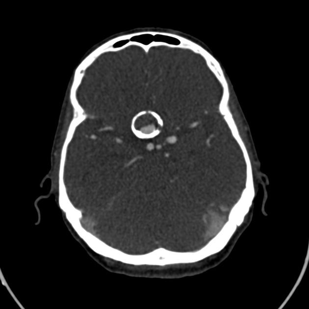
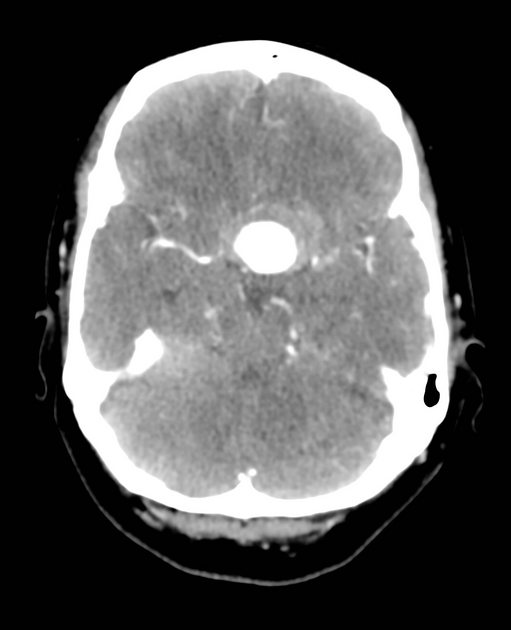
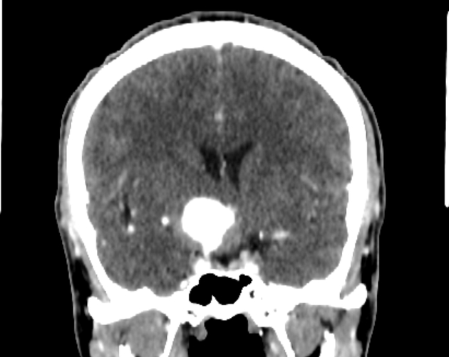
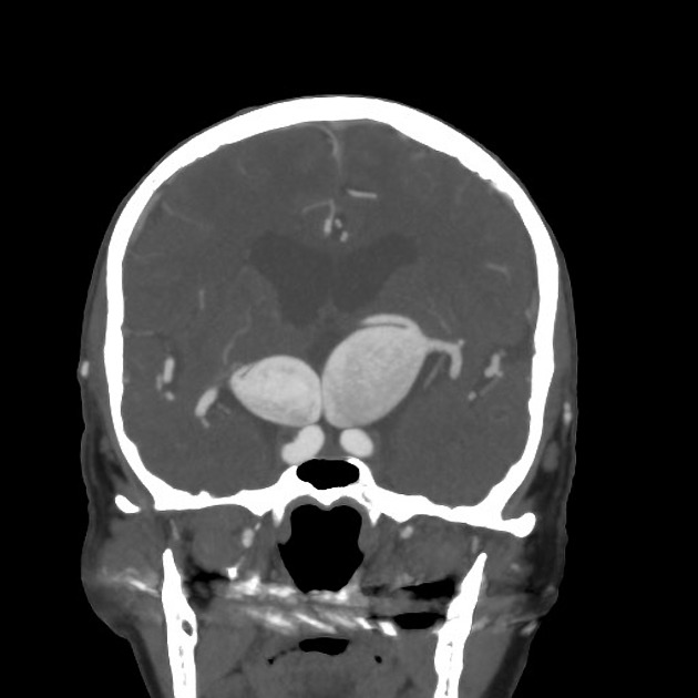
Appearances will depend on whether the aneurysm is non-thrombosed, or partially or completely thrombosed.
CT
non-contrast: slightly hyperdense, well-defined round extra-axial masses 2
may demonstrate a peripheral calcified rim
Treatment and prognosis
There are a variety of endovascular and open surgical techniques to treat these aneurysms. Endovascular options have a lower morbidity 3.


 Unable to process the form. Check for errors and try again.