
Presentation
Chronic headaches.
Patient Data
Age: 40 years
Gender: Male
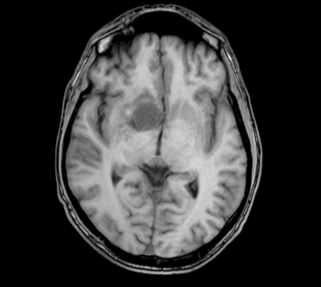
From the case:
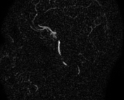
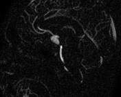
Partially thrombosed giant saccular aneurysm of the ICA

Download
Info

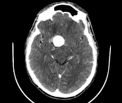
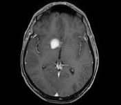
There is a well-defined homogeneously enhancing mass at the right supraclinoid region with a thick peripheral rim isodense to the cortical grey matter. No peripheral calcification is seen. No intraparenchymal, intraventricular or subarachnoid hemorrhage is seen.
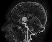
From the case:
Partially thrombosed giant saccular aneurysm of the ICA

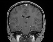
Download
Info
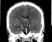
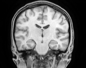
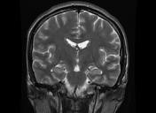
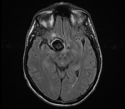
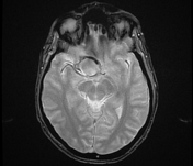
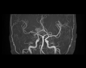
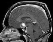
The MRI sequences demonstrate an aneurysm of the right internal carotid artery (ICA) with the following features:
- saccular type
- ovoid shape
- measures (35 x 27 x 25 mm)
- arising from the terminal segment (C7) of the ICA
- partially thrombosed. The mural hematoma is seen as an incomplete peripheral rim of high signal on T1/FLAIR and low signal on T2/T2 GE
- its lumen is of low signal on T1/FLAIR and high signal on T2 with bright and uniform enhancement on postcontrast sequences
- maximal width of the neck = 2 mm
- no apparent arterial branch taking off from the aneurysm
- mass effect is noted on the optic chiasma and 3rd ventricle
Case Discussion
CT/MRI features of a partially thrombosed giant saccular aneurysm of the ICA.
Additional contributor: A. Ramdani, MD


 Unable to process the form. Check for errors and try again.