
Hypervascular pancreatic lesions are findings that enhance more or similarly to the background pancreatic parenchyma in the late arterial phase, on contrast-enhanced CT or MRI.
Anatomical variants
- intrapancreatic accessory spleen: should not be overdiagnosed as a malignant tumour
Vascular anomalies
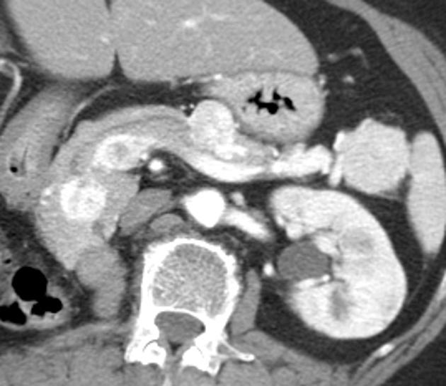
- arterial aneurysm 1
- involves the pancreaticoduodenal and /or gastroduodenal arteries
- well-circumscribed
- communicates with the adjacent vessel
- enhancement pattern identical to the aorta
- peripheral atherosclerotic calcifications may be seen
-
peripancreatic arterial pseudoaneurysm 1
- should be considered after pancreatitis or trauma
- usually surrounded by inflammation or extravasation of contrast material
- involves the splenic artery (40%), gastroduodenal artery (30%), and pancreaticoduodenal artery (10 %)
- venous aneurysm 1
- rare
- enhancement pattern identical to adjacent venous structures
- luminal communication with adjacent veins
- usually seen in the setting of cirrhosis and/or portal hypertension
- involves the splenic and portal veins
Neoplastic
- low risk of malignancy
- solid pseudopapillary tumours
- solid serous cystadenomas of the pancreas 4
- high risk of malignancy
- pancreatic acinar cell carcinoma (ACC)
- hypervascular pancreatic metastases
-
neuroendocrine tumours
- non-functional neuroendocrine tumours
- functional neuroendocrine tumours


 Unable to process the form. Check for errors and try again.